Download

1 / 46
550 likes | 1.4k Vues
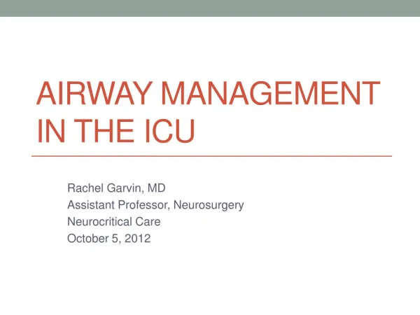
Airway Management in the Emergency Department and ICU. Global Assessment. Assess underlying need for airway control Duration of intubation - Nasal intubation less advantageous for potentially prolonged ventilator requirements Permanent support

E N D
Global Assessment • Assess underlying need for airway control • Duration of intubation - Nasal intubation less advantageous for potentially prolonged ventilator requirements • Permanent support - Underlying advanced intrinsic lung or neuromuscular disease • Temporary support • Anesthesia • Presence of reversible intrinsic lung or neuromuscular disease • Protection of the airway due to depressed mental status • Presence of reversible upper airway pathology • Patient care needs (e.g., transport, CT scan, etc.) • Significant comorbidities • Aspiration potential or increased respiratory secretions • Hemodynamic issues such as cardiac disease or sepsis • Renal or liver failure
Global Assessment • Pathophysiology of the respiratory failure • Hypoxic respiratory failure - In case of hypoxic respiratory failure, different noninvasive oxygen delivery devices can be used. - The severity of hypoxia and presence or absence of underlying disease (such as COPD) will dictate the device of choice. • Hypercapnic respiratory failure - The noninvasive device of choice for hypercapnic respiratory failure is BIPAP. • Assessment of above mentioned patient characteristics in conjunction with the mechanism of respiratory distress leads the clinician to proper choice and duration of invasive or noninvasive options for airway management. • Code status should be clarified prior to proceeding.
Global Assessment • Oxygenation • Respiratory rate and use of accessory muscles - Is the patient in respiratory distress? • Amount of supplemental oxygen - What is the patient’s oxygen demand? • Pulse oximeter or arterial blood gas - Is the patient physiologically capable of providing appropriate supply? • Airway • Anatomy - Will this patient be difficult to intubate? • Patency - Is there a reversible anatomical cause of respiratory failure as opposed to intrinsic lung dysfunction? • Airway device in place - Is there a nasopharyngeal airway or combitube in place?
Oxygen Delivery Devices(In order of degree of support) • Nasal Cannula • 4% increase in FiO2 for each 1 L of flow (e.g., 4 L flow = 37% or 6 L flow = 45%) • Face tent • At most delivers 40% at 10-15 L flow • Ventimask • Small amount of rebreathing • 8 L flow = 40%, 15 L flow = 60% • Nonrebreather mask • Attached reservoir bag allows 100% oxygen to enter mask with inlet/outlet ports to allow exhalation to escape - does not guarantee 100% delivery.
Oxygen Delivery DevicesNoninvasive Positive Pressure • CPAP is a continuous positive pressure • Indicated in hypoxic respiratory failure and obstructive sleep apnea • BiPAP allows for an inspiratory and expiratory pressure to support and improve spontaneous ventilation • Mainly indicated in hypercapnic respiratory failure and obstructive sleep apnea • If use of noninvasive modes of ventilation does not result in improved ventilation or oxygenation in two to three hours, intubation should be considered • These devices can be used if following conditions are met: • Patient is cooperative with appropriate level of consciousness • Patient does not have increased respiratory secretions or aspiration potential • Concurrent enteral feeding is contraindicated. • Facilitates early extubation, especially in COPD patients • Some devices allow respiratory rate to be set. • Up to 10 L of oxygen can be delivered into the mask for 100% oxygen delivery. • Nasal or oral (full face) mask can be used; less aspiration potential with nasal.
Degree of Respiratory Distress • Respiratory pattern • Accessory muscle use is an indication of distress. • Rate > 30 can indicate need for more support by noninvasive positive pressure or intubation • Need for artificial airway • Tongue and epiglottis fall back against posterior pharyngeal wall • Nasopharyngeal airway better tolerated • Pulse oximetry • O2 saturation less than 92% on 60 - 100% oxygen can suggest the need for intubation based on whether there is anything immediately reversible which could improve ventilation. • Arterial blood gas • pH < 7.3 can indicate need for more support by noninvasive positive pressure or intubation.
Temporizing Measures • Naloxone for narcotic overdose • 40 mcg every minute up to 200 mcg with: - 45 minutes to one hour duration of action • 0.4 - 2 mg of naloxone is indicated in patients with respiratory arrest and history suggestive of narcotic overdose - There is a potential for pulmonary edema, so large dose is reserved for known overdose and respiratory arrest • Caution in patients with history of narcotic dependence • Naloxone drip can be titrated starting at half the bolus dose used to obtain an effect - Manufacturer recommended 2 mg in 500 ml of normal saline or D5 gives 0.004 mg/ml concentration
Temporizing Measures(cont'd) • Flumazenil for benzodiazepine overdose • 0.2 mg every minute up to 1 mg • Caution in patients with history of benzodiazepine or alcohol dependence • Caution in patients with history of seizure disorder as it will decrease the seizure threshold • Artificial airway for upper airway obstruction in patients with oversedation • May be necessary in patients with sleep apnea despite judicious sedation • 100% oxygen and maintenance of spontaneous ventilation in patients with pneumothorax • Washout of nitrogen may decrease size of pneumothorax • Positive pressure may cause conversion to tension pneumothorax
Indications for Intubation • Depressed mental status • Head trauma patients with GCS 8 or less is an indication for intubation - Associated with increased intracranial pressure - Associated with need for operative intervention - Avoid hypoxemia and hypercarbia which can increase morbidity and mortality • Drug overdose patients may require 24 - 48 hours airway control. • Upper airway edema • Inhalation injuries • Ludwig’s angina • Epiglottitis
Underlying Lung Disease • Chronic obstructive lung disease • Application of controlled ventilation may interfere with complete exhalation, overdistend alveoli, and impair right heart and pulmonary venous return. • Pulmonary embolus • Pulmonary artery and right ventricle already have high pressure and dependent on preload • Application of controlled ventilation may deteriorate oxygenation and systemic pressure. • Restrictive lung disease • May require less than 6 cc/kg Vt to prevent elevated intrapulmonary pressure • Application of positive pressure may result in barotrauma in addition to impaired preload.
Airway Anatomy Suggesting Difficult Intubation • Length of upper incisors and overriding maxillary teeth • Interincisor (between front teeth) distance < 3 cm (two finger tips) • Thyromental distance < 7 cm • tip of mandible to hyoid bone (three finger breaths) • Neck extension < 35 degrees • Sternomental distance < 12.5 cm • With the head fully extended and mouth closed • Narrow palate (less than three finger breaths) • Mallampati score class III or IV • Stiff joint syndrome • About one third of diabetics characterized by short stature, joint rigidity, and tight waxy skin • Positive prayer sign with an inability to oppose fingers • No sign is foolproof to indicate intubation difficulty Prayer Sign Erden V, et al. Brit J Anesth. 2003;91:159-160.
Mallampati Score Class I: Uvula/tonsillar pillars visible Class II: Tip of uvula/pillars hidden by tongue Class III: Only soft palate visible Class IV: Only hard palate visible Den Herder, et al. Laryngoscope. 2005;115(4):735-739.
Comorbidities • Potential for aspiration requires rapid sequence intubation with cricoid pressure • Clear liquids < 4 hours • Particulate or solids < 8 hours • Acute injury with sympathetic stimulation and diabetics may have prolonged gastric emptying time. • Potential for hypotension • Cardiac dysfunction, hypovolemia, and sepsis • May need to consider awake intubation with topical anesthesia (aerosolized lidocaine) as sedation may precipitate hemodynamic compromise and even arrest. • Organ failure • Renal and hepatic failure will limit medication used. • Potential for preexisting pulmonary edema and airway bleeding from manipulation
Induction Agents • Sodium Thiopental • 3 - 5 mg/kg IV • Profound hypotension in patients with hypovolemia, histamine release, arteritis • Dose should be decreased in both renal and hepatic failure. • Etomidate • 0.1 - 0.3 mg/kg IV • Lower dose range for elderly and hypovolemic patients • Hemodynamic stability, myoclonus • Caution should be exercised as even one dose causes adrenal suppression due to similar steroid hormone structure. • Unlikely to have prolonged effect in organ failure
Induction Agents(cont'd) • Propofol • 2 - 3 mg/kg IV • Hypotension, especially in patients with systolic heart dysfunction, bradycardia, and even heart block • Unlikely to have prolonged effect in organ failure • Ketamine • 1 - 4 mg/kg IV, 5 - 10 mg/kg IM • Stimulates sympathetic nervous system • Requires atropine due to stimulated salivation and midazolam for potential of dysphoria • Avoid in patients with loss of autoregulation and closed head injury
Neuromuscular Blockers • Succinylcholine • 1 - 2 mg/kg IV, 4 mg/kg IM • Avoid in patients with malignant hyperthermia, > 24 hours out from burn or trauma injury, upper motor neuron injury, and preexisting hyperkalemia • Rocuronium • 0.6 - 1.2 mg/kg, highest dose required for rapid sequence • Hemodynamically stable, 10% renal elimination • Vecuronium • 0.1 mg/kg • Hemodynamically stable, 10% renal elimination • Cisatricurium • 0.2 mg/kg • Mild histamine release, Hoffman degradation, not prolonged in renal or hepatic failure
Rapid Sequence Intubation • Preoxygenate for three to five minutes prior to induction • Wash out nitrogen to avoid premature desaturation during intubation. • Crycoid pressure should be applied from prior to induction until confirmation of appropriate placement. • Succinylcholine 1 - 2 mg/kg IV will achieve intubation conditions in 30 seconds; Rocuronium 1.2 mg/kg IV will achieve intubation conditions in 45 seconds. • Other muscle relaxants do not produce intubation conditions in less than 60 seconds. • Avoid mask ventilation after induction. • Potentially can inflate stomach • Use only if necessary to ensure appropriate oxygenation during prolonged intubation.
Cricoid Pressure • Cricoid is circumferential cartilage • Pressure obstructs esophagus to prevent escape of gastric contents • Maintains airway patency Koziol C, et al. AORN. 2000;72(6):1018-1030.
Sniffing Position Align oral, pharyngeal, and laryngeal axes to bring epiglottis and vocal cords into view. Hirsch N, et al. Anesthesiology. 2000;93(5):1366.
Mask Ventilation • Mask ventilation crucial, especially in patients who are difficult to intubate • Sniffing position with tight mask fit optimal • May require two hands • Mask ventilation crucial, especially in patients who are difficult to intubate • Sniffing position with tight mask fit optimal • May require two hands
Laryngoscope Blades and Endotracheal Tubes Mac blade: End of blade should be placed in front of epiglottis in valecula ETT for Fastrach LMA Pediatric uncuffed ETT ETT for blind nasal Standard ETT Miller blade: End of blade should be under epiglottis
Graded Views on Intubation Grade 1: Full glottis visible Grade 2: Only posterior commissure Grade 3: Only epiglottis Grade 4: No glottis structures are visible Yarnamoto K, et al. Anesthesiology. 1997;86(2):316.
Confirmation of Placement • Direct visualization • Humidity fogging the endotracheal tube • End tidal CO2 which is maintained after > 5 breaths • Low cardiac output results in decreased delivery of CO2 • Refill in 5 seconds of self-inflating bulb at the end of the endotracheal tube • Symmetrical chest wall movement • Bilateral breath sounds • Maintenance of oxygenation by pulse oximetry • Absence of epigastric auscultation during ventilation
Additional Considerations • Always have additional personnel and an experienced provider as backup available for potential failed intubation • Always have suction available • Never give a muscle relaxant if difficult mask ventilation is demonstrated or expected • Awake intubation should be considered in the following: • If patient is so hemodynamically unstable that induction drugs cannot be tolerated (topicalize airway) • If patient has a history or an exam which suggests difficult mask ventilation and/or direct laryngoscopy
American Society of Anesthesiologists www.asahq.org
Alternative Methods • Blind nasal intubation • Bleeding may cause problems with subsequent attempts. • Contraindicated in patients with facial trauma due to cribiform plate disruption or CSF leak • Avoid in immune suppressed (i.e., bone marrow transplant) • Eschmann stylet • Fiber optic bronchoscopic intubation • Awake vs. asleep • Laryngeal mask airway • Allows ventilation while bridging to more definitive airway • Light wand • Retrograde intubation • Through cricothyrotomy • Surgical tracheostomy • Combitube
Eschman Stylet • Use especially if Grade III view achieved • Direct laryngoscopy is performed • Place Eschman where trachea is anticipated • May feel tracheal rings against stiffness of stylet • Thread 7.0 or 7.5 ETT over stylet with the laryngoscope still in place
Fiberoptic Scope • Essentially what is used to do a bronchoscopy • Can be used to thread an endotracheal tube into the trachea either while the patient is asleep or on an awake patient with a topicalized airway • Via laryngeal mask airway in place due to inability to intubate with DL: • Aintree (airway exchange catheter) can be threaded over the FOB to be placed into trachea upon visualization • Wire-guided airway exchange catheter can also be used with one more step
LMA Placement • Guide the LMA along the palate • Eventual position should be underneath the epiglottis, in front of the tracheal opening, with the tip in the esophagus • FOB placement through LMA positions in front of trachea Martin S, et al. J Trauma Injury, Infection Crit Care. 1999;47(2):352-357.
The FastrachTM Laryngeal Mask Airway • Reinforced LMA allows for passage of ETT without visualization of trachea. • 10% failure rate in experienced hands • 20% failure rate in inexperienced
The Light Wand • Transillumination of trachea with light at distal end • Trachea not visualized directly • Should not be used with tumors, trauma, or foreign bodies of upper airway • Minimal complication except for mucosal bleed • 10% failure rate on first attempt in experienced hands
Retrograde Intubation • Puncture of the cricothyroid membrane with retrograde passage of a wire to the trachea • Endotracheal tube guided endoscopically over the wire through the trachea • Catheter through the cricothyroid can be used for jet ventilation if necessary. Wesler N, et al. Acta Anaes Scan. 2004;48(4):412-416.
Emergency airway used mostly by paramedics and emergency physicians for failed endotracheal intubation Ventilation confirmed through blind blue tube Combitube is in the esophagus and salem sump can be placed through white tube Ventilation confirmed through white (clear) tube with patent distal end Combitube is in the trachea and salem sump should be placed outside of combitube into esophagus Fiber optic exchange can be accomplished through combitube Combitube
Combitube(cont'd) • Should be changed to endotracheal tube (ETT) or tracheostomy to prevent progressive airway edema • If in esophagus, take down pharyngeal cuff and attempt direct laryngoscopy (DL) or fiber optic bronchoscope (FOB) placement around combitube • Failed exchange attempt can be solved with operative tracheostomy • Placement of combitube can produce significant airway trauma • Removal prior to DL or FOB should be done with caution after thorough airway evaluation • Cricoid pressure should be maintained and emergency tracheostomy equipment available
Tracheostomy • Surgical airway through the cervical trachea • Emergent procedure carries risk of bleeding due to proximity of innominate artery • Can be difficult and time consuming in emergent situations Sharpe M, et al. Laryngoscope. 2003;113(3):530-536.
Case Scenario #1 • The patient is 70 kg with a 20-year history of diabetes. • On exam, the patient has intercisor distance of 4 cm, thyromental distance is 8 cm, neck extension is 45 degrees, and mallampati score is 1. • Your staff wants to use thiopental and pancuronium. • Do you have any further questions for this patient or would you proceed with your staff?
Case Scenario #1 - Answer • A diabetic for 20 years needs assessment for stiff joint syndrome. • You should have the patient demonstrate the prayer sign. • If the patient is unable to oppose their fingers, you should not give pancuronium. • You may want to proceed with an LMA and FOB at your disposal. • If the patient has a history of gastroparesis, you may want to consider an awake FOB.
Case Scenario #2 • 43-year-old patient with HIV, likely PCP pneumonia who had been prophylaxed with dapsone • RR is 38, oxygen saturation is 90% on 100% NRB mask • The patient is on his way to get a CT scan. • Is it appropriate to proceed without intubation?
Case Scenario #2 - Answer • Dapsone will produce some degree of methemoglobinemia. • Therefore, some degree of desaturation may not be overcome. • The patient is in significant respiratory distress and will be confined in an area without easy access. • Intubation should be considered as an extra measure of safety, especially as this patient is likely to get worse.
Case Scenario #3 • 40-year-old, 182-kg man has a history of sleep apnea and systolic ejection fraction of 25%. He has a Strep pneumonia in his left lower lobe and progressive respiratory insufficiency. • He extends his neck to 50 degrees and has a mallampati score of 2. • Would you proceed with an awake FOB?
Case Scenario #3 - Answer • The patient’s airway anatomy is not suggestive of difficulty. • However, with supine position, subcutaneous tissue may impair your ability to visualize or ventilate. • Use of gravity, including a shoulder roll, extreme sniffing position, and reverse trendelenburg may be helpful with asleep DL. • Prudent to have some accessory equipment, including an LMA and FOB, for back up
References • Caplan RA, et al. Practice guidelines for management of the difficult airway. Anesthesiology. 1993;78:597-602. • Langeron O, et al. Predictors of difficult mask ventilation.Anesthesiology. 2000;92:1229-36. • Frerk CM, et al. Predicting difficult intubation. Anaesthesia. 1991;46:1005-08. • Tse JC, et al. Predicting difficult endotracheal intubation in surgical patients scheduled for general anesthesia. Anesthesia & Analgesia. 1995;81:254-8. • Benumof JL, et al. LMA and the ASA difficult airway algorithm. Anesthesiology. 1996;84:686-99. • Reynolds S, Heffner J. Airway management of the critically ill patient. Chest. 2005;127:1397-1412.