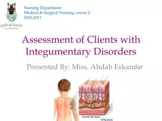
Assessing Clients with Skin Disorders
380 likes | 505 Vues
This chapter delves into the intricate functions of the integumentary system, highlighting its vital roles in protecting the body from injury, regulating temperature, and producing vitamin D. It explores the skin's layers, types of glands, and variance in skin color indicative of different health conditions. Additionally, the chapter discusses essential health assessment techniques, focusing on the physical examination and documentation of skin lesions, temperature, and turgor. Key considerations for older adults and primary and secondary skin lesions are also presented to enhance understanding for nursing professionals.

Assessing Clients with Skin Disorders
E N D
Presentation Transcript
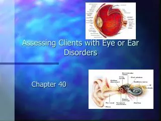
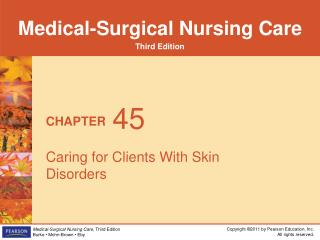
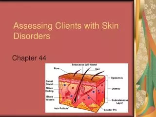
Assessing Clients with Skin Disorders Chapter 44
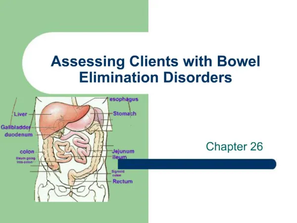
Integumentary System • Functions • 1. Protects body from injury • 2. Provides a barrier to loss of fluids • 3. Sensory - touch, pressure,pain, and temperature • 4. Regulates body temperature via sweat glands • 5. Production of vitamin D
Skin • 2 Layers • Epidermis • outer layer, protection, stores melanin • epithelial cells • Dermis • inner layer, temperature regulation • connective tissue, contains hair follicle, sweat glands and sebaceous glands
Skin Color • 1. Erythema • reddening of the skin • fever, inflammation, sunburn, drug reaction • 2. Cyanosis • bluish discoloration • poor oxygenation of hemoglobin
Skin Color • 3. Pallor • paleness of skin • shock, fear, anemia or hypoxia • 4. Jaundice • yellow-to-orange skin color • hepatic disorders
3 Types • Sebaceous - Oil • to soften and lubricate the skin • Sudoriferous - Sweat • to regulate body temperature by excretion of sweat • Ceruminous - located in external ear canal • secrete cerumen, sticky trap for foreign materials
The Hair and Nails • Protective Function • Hair • cushions the scalp • eyelashes and eyebrows protect the eyes • provides insulation in cold weather • Nails • protects fingers, toes, aid grasping
The Health Assessment Interview • Determine problems with the integumentary system • “Describe any skin problems or injuries, nail problems or scalp problems you have had.” • “Is your skin and/or scalp dry or oily?” • “Do you have any skin pain, burning or itching?”
The Physical Assessment • Can be part of head-to-toe or focused assessment • Assessment through inspection and palpation • Assess for • color, lesions, temperature,texture, moisture, turgor and edema
The Physical Assessment • Inspect color • pallor • cyanosis • jaundice • Inspect for lesions • irregular skin, rash, hives, psoriasis - scaly red patches
The Physical Assessment • Palpate the skin for temperature • warm with fever • cool in shock or decreased blood flow • Palpate skin for texture • smooth or coarse • Palpate skin for moisture • dry, moist, diaphoretic - M.I., shock
The Physical Assessment • Palpate for Turgor • pinching skin over collar bone or back of hand • decreased in dehydration tenting • increased in edema • Assess for edema • accumulation of fluid in body tissues • depress skin over ankle
The Physical Assessment • Rate the Edema • 1+ = slight pitting • 2+ = deeper pit • 3+ = obvious pit, extremities are swollen • 4+ = the pit remains • Edema occurs in cardiovascular disease, renal failure and cirrhosis of liver
The Physical Assessment • Hair • inspect distribution and quality • palpate for texture • inspect the scalp for lesions • Nails • inspect for curvature, color and thickness
Variations in the Older Adult • Loss of subcutaneous tissue • wrinkles, sagging, decreased turgor • Skin tags • small flaps of excess skin • Decreased hair and nail growth • “Liver spots” • small flat brown macules
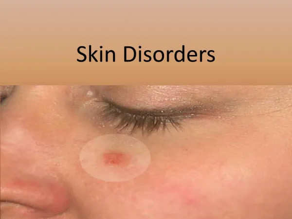
Primary Skin Lesions • Macule • flat color change in the skin - freckle • Papule • elevated palpable mass with circumscribed boarder - elevated mole • Nodule • elevated, solid mass extending deeper - lipoma
Primary Skin Lesions • Vesicle • fluid filled with thin translucent walls - blister • Wheal • larger than vesicle - insect bite, hives • Pustule • pus filled vesicle - acne • Cyst • elevated, encapsulated mass - sebaceous cyst
Secondary Skin Lesions • Atrophy • translucent, dry, paperlike skin resulting from thinning or wasting away due to loss of elastin • Ulcer • deep crater-like, irregular shaped area of skin loss extending into the dermis • Fissure • cracks with sharp edges - corner of mouth, feet
Vascular Skin Lesions • Port-wine stain • lg. Flat mass of blood vessels on skin surface • Strawberry mark • bright red, raised cluster of immature capillaries • Petechiae • flat, red-purple “freckles” caused by tiny hemorrhages
Vascular Skin Lesions • Ecchymosis • bruising - release of blood into surrounding tissues • trauma, hemophilia, liver disease • Hematoma • similar to ecchymosis but is raised, swollen
NCLEX • The nurse assessing a dark skinned client for cyanosis knows that in which of the following would cyanosis be more visible in a dark skinned individual? • A. Sclera • B. MM and nail beds • C. Generalized skin color • D. Palms of the hands and feet
NCLEX • A nurse assessing an elderly thin client notes the skin turgor over the client’s clavicle is decreased. The nurse interpretes this finding as which of the following? • A. Client is dehydrated • B. Client has edema • C. This is a normal finding for this client • D. The client has experienced a recent weight loss.
NCLEX • When performing a screening and assessment on a 44 year old female, the nurse notes a patch of hair loss. • The nurse suspects which of the following? • A. Dandruff • B. Alopecia • C. Scalp ringworm (tinea capitis) • D. head lice
NCLEX • When inspecting a client’s nails the nurse notes that the angle of the nail base is greater than 180 degrees. What is this condition called? • A. Alopecia • B. edema • C. tenting • D. clubbing
NCLEX • When working with an older person, you would keep in mind that the older adult is most likely to experience which of the following changes with aging? • A. thinning of the epidermis • B. thickening of the epidermis • C. oiliness of the skin • D. Increased elasticity of the skin
NCLEX • Which of the following glands plays a role in killing bacteria? • A. sebaceous (oil) glands • B. Eccrine sweat glands • C. Apocrine sweat glands • D. Ceruminous glands