Autoimmune Diseases and Hypersensitivity Reactions
540 likes | 602 Vues
Explore the causes of autoimmune diseases and hypersensitivity reactions, including abnormal immune responses to microbes and environmental antigens. Learn about specific diseases caused by antibodies and immune complex-mediated disorders.

Autoimmune Diseases and Hypersensitivity Reactions
E N D
Presentation Transcript
Chapter 19 Hypersensitivity Disorders(Autoimmune Diseases)
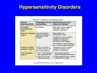
CAUSES OF HYPERSENSITIVITY DISEASES • Autoimmunity. Failure of the normal mechanisms of self-tolerance results in reactions against one’s own cells and tissues that are called autoimmunity. The diseases caused by autoimmunity are referred to as autoimmune diseases. Autoimmune diseases are estimated to affect 2% to 5% of the population in developed countries, and the incidence of these disorders is rising • Reactions against microbes. Immune responses against microbial antigens may cause disease if the reactions are excessive or the microbes are unusually persistent. Also, immune complexes, which deposit in tissues and trigger inflammation. Rarely, antibodies or T cells against a microbe may cross-react with a host tissue. Also, CTLs try to eliminate infected cells, and this normal immune response damages liver cells • Reactions against environmental antigens. Most healthy individuals do not react against common, generally harmless environmental substances, but almost 20% of the population is abnormally responsive to one or more of these substances. These individuals produce IgE antibodies that cause allergic diseases
Diseases Caused by Antibodies Against Fixed Celland Tissue Antigens
Experimental Models of Immune Complex–Mediated Diseases • Serum Sickness • Immunization of an animal such as a rabbit with a large dose of a foreign protein antigen leads to the formation of antibodies against the antigen. These antibodies bind to and form complexes with circulating antigen, which are initially cleared by macrophages in the liver and spleen. As more and more antigen-antibody complexes are formed, some of them are deposited in vascular beds • Because the complexes are deposited mainly in small arteries, renal glomeruli, and the synovia of joints, the clinical and pathologic manifestations are vasculitis, nephritis, and arthritis • More indolent and prolonged disease, called chronic serum sickness, is produced by multiple injections of antigen, which lead to the formation of smaller complexes that are deposited most often in the kidneys, arteries, and lungs
Arthus Reaction • It is induced by injection of an antigen subcutaneously into a previously immunized animal or an animal that has been given intravenous antibody specific for the antigen. Circulating antibodies rapidly bind to the injected antigen and form immune complexes that are deposited in the walls of small arteries at the injection site. This deposition gives rise to a local cutaneous vasculitis with tissue necrosis.
Systemic Lupus Erythematus (SLE) • SLE is a chronic, remitting and relapsing, multisystem autoimmune disease that affects predominantly women, with an incidence of 1 in 700 among women between the ages of 20 and 60 years (about 1 in 250 among black women) and a female-to-male ratio of 10 : 1
Clinical features • Constitutional / or from inflammation in various organ systems • Organ systems may be involved singly or in any combination • Involvement of vital organs, kidneys and CNS , accounts for significant morbidity and mortality • Morbidity and mortality result from: tissue damage due to the disease process or its therapy • Fever, anorexia, weight loss, malasia, fatigue • Skin disease or arthritis are common in SLE
The malar rash of acute lupus may be flat (malar blush) or slightly raised (butterfly eruption) and Annular rash
Renal Disease diffuse proliferative glomerulonephritis (WHO class IV) membranous glomerulonephritis (WHO class V)
Auto-antibodies in SLE The most frequent are antinuclear, particularly anti-DNA antibodies; others include antibodies against ribonucleoproteins, histones, and nucleolar antigens
Pathogenesis of SLE • Odds ratio (relative risk) for individuals with HLA-DR2 or HLA-DR3 is 2 to 3, and if both haplotypes are present, the odds ratio is about Genetic deficiencies of classical pathway complement proteins, especially C1q, C2, or C4, are seen in about 10% of patients with SLE • A polymorphism in the inhibitory Fc receptor FcγRIIB has been described in some patients • New hypotheses of the pathogenesis of SLE exposure to IFN-α, a type I interferon that is produced mainly by plasmacytoid dendritic cells and TLRs that recognize DNA and RNA, notably the DNA-recognizing TLR9, play an important role in the activation of B cells specific for self nuclear antigens • New Therapies for SLE, anti–IFN-α antibodies, inhibit TLR signals, depleting B cells by use of an antibody against the B cell surface protein CD20, blocks the B cell growth factor BAFF is now approved
Vasculitis • The systemic vascuilitis is a hetrogenous group of clinicopathologic entities characterized by inflammation and damage to the blood vessels • Most of the vasculitic syndromes are mediated by immunopathologenic mechanism • In some other forms; due to infection, malignan lymphoma, radiation, transplantation, hypersensivity play little or no role.
Immune Mediated Vasculitis • Type III Hypersensivity Polyarteritis nodosa (PAN) Henoch-Schoenlein purpura Essential Mixed Cryoglobulinemia
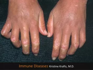
Rheumatoid Arthritis (RA) • RA is an inflammatory disease involving small and large joints of the extremities, including fingers, shoulders, elbows, knees, and ankles • Both cell-mediated and humoral immune responses may contribute to development of synovitis. CD4+ TH1 and TH17 cells, activated B lymphocytes, plasma cells, and macrophages as well as other inflammatory cells are found in the inflamed synovium, and in severe cases, well-formed lymphoid follicles with germinal centers may be present. Numerous cytokines, including IL-1, IL-8, TNF, IL-6, IL-17, and IFN-γ, have been detected in the synovial (joint) fluid • Patients frequently have circulating IgM or IgG antibodies that react with the Fc (and rarely Fab) portions of their own IgG molecules. These autoantibodies are called rheumatoid factors (RF), and their presence is used as a diagnostic test for RA. RF may participate in the formation of injurious immune complexes, but their pathogenic role is not established. Another type of antibody that has been detected in at least 70% of patients is specific for cyclic citrullinated peptides (CCP), anti- CCP
Rheumatoid Factor • Most common autoantibody in RA • Detected in 70-80% of RA patients • Bind to FC portion of IgG molecule • Usually an IgM antibody • Less often an IgA or IgG antibody • High titer predicts adverse outcome • erosive arthritis, vasculitis
Pathogenesis of RA • Susceptibility to RA is linked to the HLA-DR4 haplotype and also an association with the gene encoding a tyrosine phosphatase, PTPN22 • The identification of anti-CCP immune responses has led to new ideas about the pathogenesis of RA New Therapies for RA are antagonists against TNF, An IL-1 antagonist and an antibody against the IL-6 receptor are approved treatments, as is a fusion protein of the extracellular domain of CTLA-4 and the Fc portion of IgG, which binds to B7 molecules and blocks B7:CD28 interactions. Antibodies that block IL-17 are in clinical trials. The B cell–depleting anti-CD20 antibody is of benefit in some patients
Multiple Sclerosis and ExperimentalAutoimmune Encephalomyelitis • MS is an autoimmune disease of the CNS in which CD4+ T cells of the TH1 and TH17 subsets react against self myelin antigens, resulting in inflammation in the CNS with activation of macrophages around nerves in the brain and spinal cord, destruction of the myelin, abnormalities in nerve conduction, and neurologic deficits
MS is characterized clinically by weakness, paralysis, and ocular symptoms with exacerbations and remissions; CNS imaging suggests that in patients with active disease, there is frequent new lesion formation. The disease is modeled by experimental autoimmune encephalomyelitis (EAE) • EAE is induced by immunizing animals with antigens normally present in CNS myelin, such as myelin basic protein, proteolipid protein, and myelin oligodendrocyte glycoprotein, with an adjuvant containing heat killed mycobacterium, which is necessary to elicit a strong T cell response • EAE is caused by activated, CD4+ TH1 and TH17 cells specific for protein antigens in myelin. By analogy with the experimental disease, MS is also thought to be caused by myelin specific TH1 and TH17 cells, and these cells have been detected in patients and isolated from the blood and CNS
Pathogenesis of Multiple Sclerosis • Genetic polymorphisms associated with MS include the HLA locus, with HLA-DR2 and IL-2 receptor α chain, CD25 • Regulatory T cells are defective in MS patients • New Therapies for MS, administration of β-interferon, a random polymer of four amino acids which is postulated to bind to HLA molecules and block antigen presentation, an antibody against the VLA-4 integrin, Another recently approved drug to treat MS also interferes with leukocyte migration called fingolimod (FTY720), blocks the sphingosine 1-phosphate–mediated pathway of T cell egress from lymphoid tissues
Type 1 Diabetes Mellitus • Several mechanisms may contribute to β cell destruction, including inflammation mediated by CD4+ TH1 cells reactive with islet antigens (including insulin), CTL-mediated lysis of islet cells, local production of cytokines (TNF and IL-1) that damage islet cells, and autoantibodies against islet cells and insulin • Between 90% and 95% of Caucasians with type 1 diabetes have HLA-DR3, or DR4, or both • An informative animal model of the disease is the non-obese diabetic (NOD) mouse • New Therapies for IDDM inducing tolerance with diabetogenic peptides from islet antigens (such as insulin) or generating or giving regulatory T cells to patients.
Inflammatory Bowel Disease (IBD) • Consists of two disorders, Crohn’s disease and ulcerative colitis, in which T cell– mediated inflammation causes intestinal injury. Crohn’s disease is characterized by chronic inflammation and destruction of the intestinal wall, with frequent formation of fistulas. In ulcerative colitis, the lesions are largely confined to the mucosa and consist of ulcers with underlying foci of inflammation. New therapies for these diseases include antibodies against TNF, IL-17, and the p40 chain of IL-12 and IL-23.
Diseases Caused by Cytokine-Mediated Inflammation • In immune-mediated inflammation, TH1 and TH17 cells secrete cytokines that recruit and activate leukocytes. IL-17, produced by TH17 cells, promotes neutrophil recruitment; IFN-γ, produced by TH1 cells, activates macrophages; and TNF and chemokines, produced by T lymphocytes and other cells, are involved in the recruitment and activation of many types of leukocytes. Tissue injury results from the products of activated neutrophils and macrophages, such as lysosomal enzymes, reactive oxygen species, nitric oxide, and proinflammatory cytokines • T cell reactions against microbes and other foreign antigens may also lead to inflammation and tissue injury at the sites of infection or antigen exposure • A variety of skin diseases that result from topical exposure to chemicals and environmental antigens, called contact sensitivity, are due to inflammatory reactions. Examples of contact sensitivity include the rashes of poison ivy and poison oak (in which T cells react against chemicals made by the plants called urushiols) and rashes induced by contact with a variety of chemicals, such as thiuram, which is used in the manufacture of latex gloves
Delayed-Type Hypersensitivity (DTH) • DTH is an injurious cytokine-mediated inflammatory reaction resulting from the activation of T cells, particularly CD4+ T cells • The reaction is called hypersensitivity because it reflects excessive (i.e., injurious) immune responses (which are reflections of sensitivity to an antigen) and delayed because it typically develops during 24 to 48 hours after antigen challenge • Subsequent exposure to the same antigen (called challenge) elicits the reaction. For example, purified protein derivative (PPD), a protein antigen of Mycobacterium tuberculosis, elicits a DTH reaction, called the tuberculin reaction
Chronic DTH reactions can develop if a TH1 response to an infection activates macrophages but fails to eliminate phagocytosed microbes. If the microbes are localized in a small area, the reaction produces nodules of inflammatory tissue called granulomas