Download
1 / 25
250 likes | 527 Vues
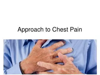
APPROACH TO CHEST PAIN Selim Krim, MD Assistant Professor Texas Tech Health Sciences Center. Objectives . Establish a differential diagnosis for chest pain Know what clues to obtain on history to rule-in or out MI, PE, pneumothorax and aortic dissection Identify risk factors for MI

E N D
APPROACHTOCHEST PAIN Selim Krim, MD Assistant Professor Texas Tech Health Sciences Center
Objectives • Establish a differential diagnosis for chest pain • Know what clues to obtain on history to rule-in or out MI, PE, pneumothorax and aortic dissection • Identify risk factors for MI • Know how to do a focused physical exam, identifying features that would distinguish between MI, PE, pneumothorax and aortic dissection. • Identify investigations required in diagnosing MI • Outline management strategy in MI
Etiologies • Myocardial ischemia or infarction • Pulmonary embolus • Pneumothorax • Pericarditis • Tamponade • Pneumonia • Aortic dissection • Gastritis, peptic ulcer disease • Musculo-skeletal • Shingles
As a general rule any chest pain is ischemic in origin until proven otherwise!
Myocardial ischemia or infarction • Pressure-type of chest pain • Generally involves central to left-sided pain with radiation to jaw or arms • Exacerbated by activity, relieved with rest • Relieved with nitro spray • Associated with nausea, diaphoresis, syncope, shortness of breath • Enquire about cardiac risk factors: age, sex, smoking history, diabetes, hypertension, hyperlipidemia, previous myocardial infarction and family history
Myocardial ischemia or infarction • ↓BP indicates cardiogenic shock • ↑JVP, pulsatile liver and peripheral edema seen in right-sided heart failure • Oxygen desaturation, crackles, S3 seen in left-sided heart failure • New murmurs: mitral regurgitation murmur in papillary muscle dysfunction
Work-up • EKG (should be knee-jerk reflex in chest pain scenario!) • CXR to look for signs of congestive heart failure • Cardiac enzymes: CK (will begin to rise 6 hours after infarct and remain elevated for 24-48 hours), troponin (will begin to rise 12 hours after infarct and remain elevated for 2 weeks). Need to follow serially if first set negative.
Management Strategy for NSTEMI Initial therapy • Morphine for pain • Oxygen if hypoxic • Nitro spray/drip for pain • Aspirin
Management Strategy for NSTEMI/NST Chest Pain • Establish risk level using the TIMI scoring system: • Low risk: May be discharged after symptom control • Moderate risk: Admit for further evaluation; add beta blockers , Ace inhibitors . Follow cardiac enzyme levels. If Mi ruled out, Exercise or Adenosine stress test before discharge • High Risk: Admit for cardiac catheterization
Management Strategy for STEMI • Morphine, oxygen, nitro, aspirin • Beta blockers, Ace inhibitors • Early invasive strategy with either thrombolytic therapy or percutaneous coronary intervention (preferred)
Pulmonary Embolism • Sudden-onset sharp chest pain • Exacerbated by inspiratory effort • Can be associated with hemoptysis, sycope, dyspnea, calf swelling/pain from DVT • Risk factors: immobilization, fracture of a limb, post-operative complications, hypercoagulable states (underlying carcinoma, high-dose exogenous estrogen administration, pregnancy, inherited deficiencies of antithrombin III, activated protein C, S, lupus anticoagulant, prior history of DVT/PE [Virchow’s triad]
Pulmonary Embolism • Anxious patient, sense of impending doom • Tachycardia, tachypnea, hypoxia • EKG: sinus tachycardia most common, S1Q3invertedT3 with large embolus (classic, but rare!), look for right-axis deviation • V/Q scan very sensitive but not specific • Spiral CT with contrast show large, central emboli • Pulmonary angiogram is gold standard but carries risk • Consider Doppler U/S of legs
Pneumothorax • Can be asymptomatic or present with acute pleuritic chest pain and dyspnea • Primary pneumothorax predominantly in healthy young tall males • Due to trauma (MVA accidents – associated with rib fractures, iatrogenic – during line placement, thoracentesis) • Increased alveolar pressure from asthma or barotraumas (BiPAP, ventilator-associated) • Rupture of bleb in COPD patients
Pneumothorax • Decreased expansion of chest • Decreased breath sounds and • Decreased tactile/vocal fremitus on side of pneumothorax • Hyperresonant percussion note • Usually easily confirmed by CXR
Aortic Dissection • Abrupt onset • The pain usually is described as ripping or tearing • Tearing or ripping pain that is felt in the intrascapular area • New diastolic murmur, asymmetrical pulses, and asymmetrical blood pressure measurements • Risk factors: HTN, Marfan syndrome, coarctation of aorta.. • Widened mediastinum on a portable anteroposterior (AP) radiograph • TEE considered diagnostic test of choice
Case 1 A 64-year-old woman is evaluated in the emergency department 6 hours after the onset of severe crushing chest pain associated with diaphoresis, nausea, and vomiting. Her medical history is significant only for mild hyperlipidemia. Her medications include atorvastatin and aspirin. Her blood pressure is 150/88 mm Hg, and her pulse rate is 88/min. The lungs are clear; she has no murmurs; examination of the abdomen and extremities is normal. What is the best next step in the management of this patient? • CXR • EKG • Cardiac enzymes • CBC
Case 1 Electrocardiogram shows a 3-mm ST-segment elevation in leads II, III, and aVF, with occasional premature ventricular contractions. Cardiac enzymes are elevated. What is the next step in the management of this patient? • Thrombolytic therapy • Coronary angiogram • Beta blockers • Amiodarone
Case 2 A 72-year-old man is evaluated in the emergency department for the sudden onset of severe sharp anterior chest pain radiating into the back. He is a former smoker with a long history of type 2 diabetes mellitus, chronic renal insufficiency (creatinine 2.0 mg/dL [176.84 μmol/L]), sick sinus syndrome with a DDD pacemaker implanted in 1995, and hypertension. His medications include insulin, furosemide, ramipril, and aspirin.
Case 2 On examination, the blood pressure is 185/85 mm Hg bilaterally, and the pulse rate is 90/min and regular. A grade 2/6 systolic murmur and a soft decrescendo diastolic murmur are heard at the second right intercostal space. There are abdominal and bilateral femoral bruits, with absent distal pulses. His EKG shows no ST, T wave changes. CXR is normal. Which of the following is the most appropriate initial imaging study? • Non-contrast chest CT • Chest MRI • Transesophageal echocardiography • Transthoracic echocardiography
Case 3 • A 64-year-old man is evaluated in the emergency department for epigastric chest discomfort and episodes of dyspnea with moderate activity. The discomfort started 2 days ago and has been intermittent, occurring mostly at rest. He works in an office and is relatively inactive. He had been using antacids for several months with variable response. He has no significant medical history and takes no other medications.
Case 3 Blood pressure is 150/85 mm Hg and heart rate is 81/min; there is no jugular vein distention or carotid bruits; cardiac examination reveals a normal S1 and S2, with no murmur, gallop, or clicks. Examination of the abdomen and extremities is normal. Electrocardiogram shows flattened T waves. What is your next step in the management of this patient? • Discharge him home with follow up with his physician • Admit to rule out Myocardial infarction • Emergent coronary angiogram • Thrombolytic therapy
Key Points • Not every chest pain is MI, however every chest pain should be considered as ischemic until proven otherwise • A good history and physical exam may help with the diagnosis • EKG is the best single diagnostic test to help rule out MI • Use the TIMI scoring system to help for the diagnosis and prognosis of MI