Landmarks for Inguinal Ligament
430 likes | 1.93k Vues
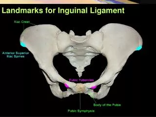
Landmarks for Inguinal Ligament. Inguinal region. Boundaries Inguinal ligament Lateral margin of rectus abdominis A horizontal line stretching from anterior iliac spine to lateral margin of rectus abdominis. Inguinal region - Layers. Skin Superficial layer Camper’s Scarpa’s.

Landmarks for Inguinal Ligament
E N D
Presentation Transcript
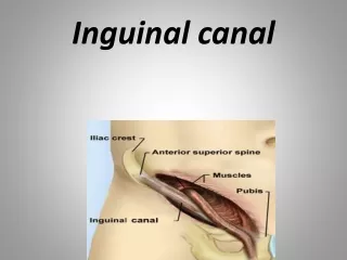
Inguinal region Boundaries • Inguinal ligament • Lateral margin of rectus abdominis • A horizontal line stretching from anterior iliac spine to lateral margin of rectus abdominis
Inguinal region-Layers • Skin • Superficial layer • Camper’s • Scarpa’s
Inguinal canal Position: oblique passage, 4cm long, located 1.5cm above medial half of inguinal ligament
Boundaries of inguinal canal • Anterior wall • Aponeurosis of obliquus externus abdominis • Obliquus internus abdominis (lateral third of wall) • Posterior wall • Transverse fascia • Inguinal flax medially • Roof-arching fibers of obliquus internus and transversus abdominis • Floor-inguinal ligament
Boundaries of inguinal canal • Roof • internal oblique abdominis • transversusabdominis • Floor • inguinal ligament • lacunar ligament • Anterior wall • external abdominal oblique aponeurosis • internal abdominal oblique aponeurosis • Posterior wall • transversalis fascia • conjoint tendon (falxinguinalis)
Boundaries of inguinal canal Two openings • Superficial inguinal ring-a triangular-shaped defect in the aponeurosis of the obliquus externus abdominis and lies immediately above and medial to the pubic tubercle
Boundaries of inguinal canal Two openings • Deep inguinal ring-an oval opening in transverse fascia and lies approximately 1.5cm above midpoint of inguinal ligament
Mechanics of the inguinal canal • The inguinal canal is a site of potential weakness in both sexes. • On coughing and straining (as in micturition, defecation, and parturition), the arching lowers fibers of the obliquusinternus and transversusabdominis contract and flatten the arch. • In turn, this lowers the roof of the canal toward the floor and virtually closes the canal.
Structures passing through the inguinal canal • In males • Spermatic cord • Ilioinguinal nerve • In females • Round ligament of uterus • Ilioinguinal nerve
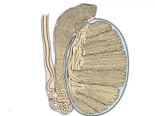
Structures passing through the inguinal canal-Male • Spermatic cord • arteries: testicular artery, deferential artery, cremasteric artery • nerves: genital branch of the genitofemoral nerve, nerve to cremaster, sympathetic nerves • vas deferens • pampiniform plexus • lymphatic vessels • Ilioinguinal nerve
Structures passing through the inguinal canal-Females • Round ligament of the uterus • Ilioinguinal nerve
Descent of testes Seven-week embryo showing the testis before its descent from the dorsal abdominal wall
Descent of testes Fetus at 28 week the testis passing through the inguinal canal
Descent of testes 4 months 11 weeks 8 months
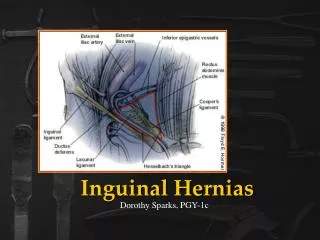
Inguinal Triangle (of Hesselbach) Boundaries • Medially: Lateral border of rectus abdominis • Laterally: Inferior epigastric artery • Inferiorly: Inguinal ligament
Layers-External Oblique Abdominis Inguinal Lig. Lacunar Lig. Pectineal Lig. (Cooper’s Lig.)
Inguinal Ligament Reflected ligament Intercrural fibers Lateral crus Medial crus Superficial inguinal ring
InguinalLigament EOA EOA EOA Inferior epigastric vessels Cremaster Conjoint Tendon
InguinalLigament Transverse abdominalfascia Abdominal inguinal ring (deep inguinal ring)
Anterior Abdominal Wall: Internal View • Extraperitoneal fascia • Parietal peritoneum • Medianumbilicalfold(medianumbilicalligamentformedbyobliteratedurachus) • Medialumbilicalfold(medialumbilicalligamentformedbyobliteratedumbilicalartery) • Lateralumbilicalfold(Inferiorepigastricvessels)
Anterior Abdominal Wall: Internal View • Supravesical fossa • Medial inguinal fossa • lateral inguinal fossa
Inguinal Hernia • A hernia is the protrusion of part of the abdominal contents beyond the normal confines of the abdominal wall • Consists of three parts: the sac, contents of the sac, covering of the sac • Hernial coverings are formed from the layers of the abdominal wall through which the hernial sac passes
Indirect Inguinal Hernia • It is the most common form of hernia • Is believed to be congenital in origin • The hernial sac is remains of processus vaginalis • Enters the inguinal canal through the deep inguinal ring lateral to the inferior epigastric vessels • It may extend part of the way along the canal or as far as the superficial inguinal ring
Indirect Inguinal Hernia • If the processus vaginalis has undergone no obliteration, the hernia is complete and extends through the superficial inguinal ring down into the scrotum or labium majus • Under these circumstances the neck of the hernial sac lies at the deep inguinal ring • It is 20 times more common in young males than females • Is more common on the right side