Lecture 5
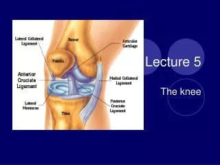
Lecture 5. The knee. Anatomy Review Knee joint Particularly susceptible to injury because it is located between two long levers (Tibia and Femur) Depends on ligaments and muscles for stability not bony configuration. Tibiofemoral joint Modified hinge joint – 3 degrees of freedom

Lecture 5
E N D
Presentation Transcript
Lecture 5 The knee
Anatomy Review • Knee joint • Particularly susceptible to injury because it is located between two long levers (Tibia and Femur) • Depends on ligaments and muscles for stability not bony configuration
Tibiofemoral joint • Modified hinge joint – 3 degrees of freedom • Synovial membrane • Resting position 25 degrees of flexion
Menisci (discs of fibrocartilage) • Medial – C shaped thicker posteriorly • Lateral – O shaped equal thickness • Both are thicker along the periphery and thinner along the inner margin • Both move posteriorly during movement from extension to flexion • Both are avascular in the inner two thirds and partly vascular in the outer third
Both attach in the middle of the tibia on the intercondyler notch • Medial is held in place by the coronary ligament (attached to tibia on medial edge) • Lateral is not attached to the outside of the tibia – therefore less likely to be injured
Functions of the Menisci • aid in lubrication of the joint • acts as shock absorbers – spreads out stresses • makes joint surfaces more congruent • improves weight distribution by increasing contact area • reduces friction • helps to prevent hyperextension
Minimal or no pain when damage menisci – due to lack of innervation • Minimal effusion – due to lack of blood supply – however usually damage synovial membrane as well and this increases swelling • Low potential for healing due to avasucular in nature – especially inner two thirds
Patellofemoral Joint • modified plane joint • patella thickest layer of cartilage in the body • 3 distinct facets • medial • lateral • each having superior , middle and inferior surfaces • odd lying medial to medial facet • Odd first to be effected in PFS • Odd comes into contact with femur at 135 degrees of flexion – deep squats
Patella • improves efficiency of extensors especially during last 30 degrees of extension • acts as a guide for quads tendon • decreases friction of quads mechanism • bony shield
Patella Loading • Walking 0.3 X BW • Climbing stairs 2.5 X BW • Descending stairs 3.5 X BW • Squatting 7 X BW
Ligaments • MCL • Two layers - deep and superficial • Deep is a thickening of the capsule blends with medial meniscus • Superficial strong rectangular shaped band • Runs from medial epicondyle to medial surface of tibia • MCL is tight through out the ROM
LCL • round and lies under the biceps femoris tendon • runs from lateral epicondyle to the head of the fibula • tight in extension • loose in flexion • separate from the capsule and meniscus
Cruciates • ACL & PCL • cross each other • primary rotary stabilizers of the knee • Intracapsular (with articular capsule) but extra synovial (lie outside synovial cavity) • Named in relation to attachment on the tibia
ACL • Prevents anterior translation of the tibia on the femur • Checks lateral rotation of the tibia in flexion • Helps control the normal rolling and gliding of the knee joint • Least amount of tension on ACL between 30 –60 degrees of flexion • Taut in both flexion and extension
PCL ( shorter and stronger than ACL) • Primary stabilizer of the knee against posterior translation of tibia on femur • Checks extension and hyperextension • Bulk of fibers tight at 30 degrees flexion
ROM of Knee / End Feel • Flexion/Extension • Muscles?
Valgus • MCL • Athlete is supine or high sitting • One plane medial instability test • Examiner pushes the knee medially with heel of hand, other hand stabilizes distally • Tested in full extension - all fibers • Tested in 20 –30 degrees of flexion – superficial fibers • Positive test – pain and gapping
Varus • LCL • Athlete is supine or high sitting • One plane lateral instability test • Examiner pushes the knee laterally • Usually done in slight flexion • Positive test – pain and lateral gapping • http://www.youtube.com/watch?v=ubP-1WaFeEc&feature=related
Anterior Drawer Test • ACL • athlete supine - hip flexed to 45 and knee to 90 degrees • foot (in a neutral position) stabilized by examiner sitting on it • place both thumbs on either side of patella tendon , fingers in popliteal fossa (ensure hamstrings are relaxed)
while palpating the joint line apply an anterior displacement on the tibia • positive test-anterior translation of tibia on femur
Lachmans • ACL • Athlete is in supine or high sitting • Leg is between 20 to 30 degrees of flexion • Examiner stabilizers the femur with one hand and tries to pull tibia forward on the femur with other hand • Positive test - forward translation of tibia on femur (soft mushy feeling)
Modified Lachman’s (Drop Lachman’s) • Often used by people with small hands • weight of the femur is supported by the table • http://www.youtube.com/watch?v=VqekkznP-Lw&feature=related • http://www.youtube.com/watch?v=dH_jnTy1rNk&feature=related
Posterior Sag Test • PCL • Athlete is supine or high sitting • Knees flexed to 90 degrees • Hip flexed to 40 degrees (or 90 ) • Heel lined up with each other • Examiner looks across side of knee to see if tibial tuberosity's are equal • Positive test - is a sag of one tibia on the femur • http://www.youtube.com/watch?v=i-nFGqwr_VE&feature=related
PCL and ACL Tear • http://www.youtube.com/watch?v=lVaZ710s804&feature=related
McMurry’s • Menisci • Athlete in supine or high sitting • Knee flexed to chest and tibia is rotated medial and extend hip and knee, repeat with lateral rotation • Positive test- click , pop or pain • Medial rotation – lateral meniscus • Lateral rotation – medial meniscus • http://www.youtube.com/watch?v=fkt1TOn1UfI&feature=related
Bounce Home • Fat pad or menisci • Athlete is supine • Knee is flexed by examiner and then allowed to drop into extension • Positive test – springy block , or not able to completely extend the knee (menisci) • NO pain and just a block may be a fat pad or bursa
Tests for swelling • Brush / Stroke test • Examiner brushes up the medial side of the knee and down the lateral side • Looking for fluid to move from one side to the other • http://www.youtube.com/watch?v=N0nXJB3aQBk • Patellar tap test • Slight pressure is placed on the patella • If positive the patella feels like it is floating
Clark’s Sign • PFS • Athlete is supine or high sitting • Examiner pushes distally on the top of the patella while the athlete slowly contracts their quads • Positive test- if there is pain – however this is painful for most individuals – not a great test • http://www.youtube.com/watch?v=NY5I0HlxoFE&feature=related
Patella Apprehension Test • subluxing patella • With the knee relaxed the examiner pushes the patella laterally • Positive test - If the athlete shows apprehension • http://www.youtube.com/watch?v=xXmjYVDkmVg&feature=related
Nobel Compression test • ITB • Athlete is sitting with legs hanging over the edge of a table • Pressure is applied to the ITB just above the knee • The athlete’s leg is then extended either actively or passively • Positive test -is pain during the last 30 degrees or so of extension or clicking or snapping.
Q- Angle • angle between the RF and the patellar tendon • Measure from ASIS to mid point of the patella • And from tibial tuberosity to mid point of the patella • Q-angle is formed by these two intersecting lines
Males – 13 degrees (10 – 15) • Females 18 degrees (10 - 19) • The greater the Q- angle the more likely the athlete will have problems with PFS
Reflexes • Patella (L4,L5) • Achilles ( S1)
Muscle testing • Knee Extension • Quads – RF, VM, VL, VI. • Athlete is high sitting with leg over edge of table flexed to 90 degrees • Examiner applies resistance to lower leg just above ankle and asks athlete to try to extend the leg • Athlete must meet the resistance of the examiner, do not let them overpower you • Stabilize at the thigh • Positive test- pain and weakness
Knee Flexion • Hamstrings – SM, ST, BF • Athlete is prone • Hip is in neutral and leg is flexed to 90 degrees • Resistance is applied by the examiner trying to extend the leg • Thigh is stabilized • Positive test- pain and weakness
Sport Specific Functional Tests • forward running • cross-over running • figure 8’s • v- cuts • side steps • karioca running
![[lecture#5]](https://cdn0.slideserve.com/109460/slide1-dt.jpg)