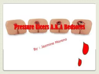
Pressure Ulcers
Pressure Ulcers. Jennifer E. Marks, D.O. February 25, 2004 LRI. Definition of Pressure Ulcer. An area of unrelieved pressure over a defined area, usually over a bony prominence such as the sacrum

Pressure Ulcers
E N D
Presentation Transcript
Pressure Ulcers Jennifer E. Marks, D.O. February 25, 2004 LRI
Definitionof Pressure Ulcer • An area of unrelieved pressure over a defined area, usually over a bony prominence such as the sacrum • Pressure leads to ischemia, cell death, and tissue necrosis, as capillaries are compressed and the blood flow is restricted • Muscle is the most sensitive tissue to pressure, skin is the most resistant
How do pressure ulcers usually present? • 1. High pressure over bony prominence (can be a single insult) • 2. At muscle and bone interface, ischemia results. • 3. Affected area with erythema, induration, warmth, and skin is intact • 4.Days to weeks s/p insult, EVEN WITH PRESSURE RELIEF, the wound opens, and is a depression with necrotic tissue
Pressure ulcers • Associated with : • 1. Shear forces • 2. Impaired sensorium/sensation (SCI patients!) • 3. Poor nutrition- serum albumin positively correlates with pressure ulcer stage, and negatively correlates with risk • 4. Chronic illness • 5. Elevated tissue temperature- Higher metabolic demands • 6. Maceration
SCI patients • Increased risk for pressure ulcer formation • Estimated incidence 25-66%
Pathomechanics • Shear forces- Tangential to the skin surface. Can play a major role in the formation of sacral ulcers. • Axial forces- Perpendicular to the skin surface. Unrelieved axial pressure 4-6 times the systolic pressure can cause necrosis in less than 60 minutes! • If tissue capillary pressure is exceeded , ulcers will form at that site.
How much pressure is too much? • Kosiak 1961- Studied the effects of pressure and time on rat muscle. • More pressure=less time for an ulcer to form • Found that alternating pressure of as little as 5 minute intervals led to considerably less ulcer potential.
Kosiak’s research led to the current practice of turning patients every two hours. • Why don’t we measure pressure/shear as clinicians? • Transducers are thick , bulky, and expensive • Shear transducers have not been modified for clinical use
What is a safe amount of pressure? • Studies by Landis et al. have led clinicians to believe that pressures under 32 mm Hg are generally believed safe • This value is influenced by tissue stiffness, tissue composition, and the patient’s body contour
Clinical Wound Assessment • 1. Color photography- Use ruler in picture to give dimensions. Very useful when done in a serial fashion. An alternative is to draw pictures of the wound. • 2.Location- Be specific. • 3.Size- Be sure to include length, width, and depth measurements, in centimeters.
Clinical Wound Assessment • 4. Describe the type of irrigation utilized, and the dressing type. • 5.Drainage: • Amount(minimal, moderate, copious) • Color (serous, serosanguinous, prurulent) • Odor(present, absent)
Clinical Wound Assessment • 6. Undermining/tunneling – Present/absent • 7. Wound character- What kind of tissue? Is there granulation, slough? • Stage the ulcer • IF THERE IS ESCHAR PRESENT, YOU CANNOT STAGE THE WOUND!
Treatment • Sharp debridement • Mehanical nonselective debridement • Enzymatic Debridement • Autolytic debridement
Sharp debridement • Removal of devitalized tissue/eschar via surgical means. Small wounds can be debrided at bedside, more extensive wounds can be addressed in the OR • Most effective/quickest method of removing necrotic tissue. Debridement is done to the point where the tissue bleeds with forceps and a scalpel. • Must have the clinical skill/judgement necessary to be able to discern the difference between vitalized and nonvital tissue. • Cons: Can damage healthy tissue
Mechanical nonselective debridement • Whirlpool • Irrigation • Wet to Dry dressing- utilize normal saline, place moist gauze on the wound, let dry. • When the dressing is removed, the necrotic tissue comes off it. • Cons: Healthy tissue can be damaged
Wet to Dry vs. Wet to Moist • Saline wet to dry dressings are used to debride necrotic wounds • Wet to moist dressings maintain a clean moist wound bed and are removed before they dry out. • Wet to moist dressings have to be changed more frequently
Enzymatic Debridement • Utilizes chemical agents (such as Accuzyme) in the form of ointments which work on the necrotic wound debris, and do not affect the viable tissue
Autolytic debridement • The bodies own enzymes break down dead tissue.Wound cells secrete proteases, collagenases that digest eschar. Hydrocolloid dressings help to promote this type of debridement. • Pros: Very effective in noninfected wounds- an occlusive dressing allows wound fluid to collect • Cons: If the wound is infected, you have just created an abcess!
Wound dressings • Gauze • Transparent adhesive dressings • Hydrocolloid dressings • Gel dressing • Calcium alginate dressings
Transparent adhesive dressings • Ex. Tegaderm, Opsite • Semipermeable, occlusive • Stage I/II wounds without debris • Allow gaseous exchange/water vapor transfer from the skin, prevent peri-wound maceration • Do not use if wound is exudative or the patient is diaphoretic
Hydrocolloid dressings • Ex. Duoderm • Interact with wound exudate , and make a gel • Keep wound surface moist. • Enhances healing, protects versus secondary infection • Help to minimize shear • Good for shallow stage III sacral ulcers
Calcium alginate dressings • Made from brown seaweed (ex. Sorbsan), sterile • Semi-occlusive, highly absorbable • Good for treating exudative or contaminated wounds • Need to be frequently changed
Most common pressure ulcer sites • Ischium 28% • Sacrum 17-27% • Trochanter 12-19% (Bears weight when patient is in a sitting position) • Other commonly affected sites include coccyx, heel, and malleolus
Treatment • Proper medical care • Turn patient Q 2 hours • Frequent dressing changes • Proper nutrition- High protein diet indicated as a high amount of protein is lost through the wound • Pressure relief in wheelchair, specialty support surfaces • Continued wound assessment
Support surfaces • Include overlays (water, gel , foam, air) • Specialty beds • Low air loss beds (Flexicare)have cushions filled with air that keep pressures below the capillary closing pressures • Air fluidized beds (Clinitron) use warm air forced through silicone beads to mimic a fluid medium
Which support surface is best? • No study has shown conclusively that one surface performs better than the others! • Must individualize your approach • If a patient has a Stage III or IV ulcer, the patient should be utilizing a pressure relief product
Wound Infection • Presentation: Foul odor, greenish drainage, dull white base (versus red granulation tissue). Can have cellulitis, with erythema, warmth, swelling, tenderness. • Systemic bacteremia: Chills, anorexia, nausea/vomiting, fever, increased white count, mental status changes, glucose intolerance in diabetics. • Signs of bacteremia/cellulitis- IV abx/possible debridement
Wound cultures • Should not be routinely performed, as the cultures will always be positive • Exception- If antiseptic such as Betadine is used prior to local debridement, and an abcess or other sequestered collection is exposed • Occasionally, cultures are taken for burn wounds • Greater than 105 CFU’s- wound will not heal
When are topical antibiotics indicated for pressure ulcers? • If a pressure ulcer does not heal after 2-4 weeks of optimal treatment, can try silver sulfadiazine or triple antibiotic ointment x 2-3 weeks
Osteomyelitis • Must keep in mind , especially with a Stage IV pressure ulcer or if ulcer over a bony prominence • 25% of nonhealing ulcers have bone infection • Gold standard- Bone biopsy • Imaging- XRay, MRI
Xray • Reactive bone formation and periosteal elevation =osteomyelitis • BONE SCANS ARE A POOR STUDY TO DETECT OSTEOMYELITIS! High false positive rate.
MRI • 95% sensitive • On T2 weighted image, can demonstrate marrow edema • Can reveal soft tissue abnormalities such as perirectal fistulas
Why don’t we just close the wound (vs. using flap)? • Usually not enough soft tissue • Too much tension where the incision site would be
Musculocutaneous flaps • Why use a muscle flap when muscle is the first tissue to become ischemic? • Minimizes “deadspace”, and provide tissue with rich vasculature • NOT being used as a cushion! • In less than a year, the flap usually atrophies, but the blood supply remains intact • Also, the surgeon can put the suture line away from the maximal pressure area
When are flaps indicated? • Better for an SCI patient , or when muscle loss will not affect ambulatory ability • Not as easy a decision in an ambulatory patient, where function can be compromised
Best treatment for a pressure ulcer? • NO ONE RIGHT ANSWER! Keep in mind: • 1. PREVENTION • 2. Must correct the underlying problem • 3. Wounds must be cleaned/ dead tissue removed before healing can occur • 4. Keep wound moist- Permits cells to perform migration/mitosis
What is being researched? • Electrical stimulation – some studies are showing improved pressure ulcer healing rate in chronic stage III and IV ulcers • Controversial • Also small study using growth factors in SCI patients. • Both areas need more research
Works Cited Braddom, Randall L. Physical Medicine and Rehabilitation. Second Edition. Philadelphia, Pennsylvania. W.B. Saunders Company, 2000. O’Young, Young, et al. Physical Medicine and Rehabilitation Secrets. Second Edition. Philadelphia, PA. Hanley & Belfus Inc., 2002.