Download
1 / 29
290 likes | 630 Vues
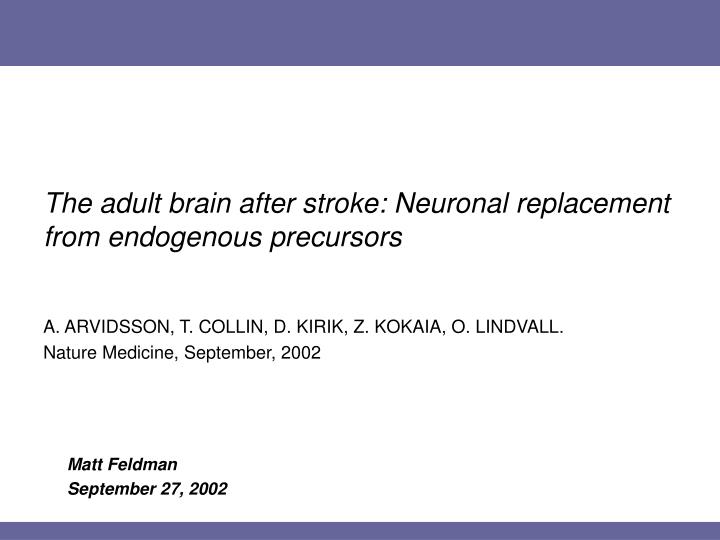
The adult brain after stroke: Neuronal replacement from endogenous precursors. Matt Feldman September 27, 2002. A. ARVIDSSON, T. COLLIN, D. KIRIK, Z. KOKAIA, O. LINDVALL. Nature Medicine, September, 2002. Overview. Background: neurogenesis Up to here: previous research

E N D
The adult brain after stroke: Neuronal replacement from endogenous precursors Matt Feldman September 27, 2002 A. ARVIDSSON, T. COLLIN, D. KIRIK, Z. KOKAIA, O. LINDVALL. Nature Medicine, September, 2002
Overview • Background: neurogenesis • Up to here: previous research • This work: what’s involved and why • The system of study • Techniques and markers for measuring proliferation • The question • Is true neurogenesis observed? • The implications and the future
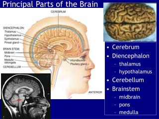
Background • The adult brain: relevant anatomy Neurogenesis observed in the SVZ, dentate gyrus and olfactory bulb 1. Ventricle and subventricular zone (SVZ) 2. Striatum
Background: Magavi et al. • Magavi et al. (Nature 405, 951–955 (2000)) • induced neuronal degradation and examined fate of dividing cells • chromophore-targeted apoptosis of pyramidal neurons of the cortex induced neurogenesis some reconstitution of damaged area • 3D laser scanning confocal microscopy confirmed that new cells are not merely closely in close proximity to pre-existing neurons • pyramidal morphology indicative of long distance projections • additional labeling was negative for GFAP and MBP (immature markers) new neurons had fully differentiated
Background: Magavi et al. • Cell division can continue after an injury • But, unlike a clinical stroke event, lesion only affected targeted neurons • Method makes damaged/destroyed neurons the source of the injury, rather than the pathological outcome • Ignores tissue complexity • All the surounding cells (and precursors they express) are still intact • Relatively small lesion • Quiescent, but pre-determined survivors may differentiate with signals from adjacent cells
Approaching clinical relevancy • Neurogenesis is observed in the adult brain • After a more clinically relevant event (ischemic stroke – localized anemia following occlusion), is similar neurogenesis observed? • Can new neurons migrate to the site of an injury? • If so, are they appropriate? Long-lived? Are endogenous precursors sufficient to stimulate neurogenesis in adult rat striatum following stroke?
Methods: MCAO • Injury model employed middle cerebral artery occlusion (MCAO) technique • monofilament inserted into common carotid artery and advanced to middle cerebral artery, held for 2 hours • Sham: filament placed into common carotid, no forward advancement
Methods: Markers of proliferation 5-bromo-2’-deoxyuridine (BrdU) • Newly-injected BrdU is available for a few hours for incorporation • Replaces tritiated thymidine and autoradiographic assays with immunological quantification • Fluorescent Ab tagging in multiple excitation channels allows for simultaneous measurement of different probes • DNA synthesis/cell proliferation measured by BrdU incorporation during S phase; detection using anti-BrdU monoclonal Antibody
Methods: Markers of neurogenesis Neuronal nuclear antigen (NeuN) • Neuron-specific nuclear protein (vs cytoplasmic or cell-surface antigen) observed in invertebrates • Recognized with a mAb in standard IHC • Specifically reactive for post-migratory (late maturity) neurons • No non-specific (ex. glial) reaction within NS; no non-neuronal detection • Doesn’t detect all types of neurons, but most
Stroke leads to neurogenesis in damaged striatum Individual neuron in X-Y plane NeuN BrdU NeuN/BrdU Successive sections of neurons in the Z plane
Stroke leads to neurogenesis in damaged striatum 137 Cell number Cell density 29 0.8 • BrdU injected 2x/day during days 4,5,6 post-stroke (n=9; 10) • 31-fold increase in number of BrdU/NeuN-labeled cells • Few observed BrdU/NeuN cells in contralateral striatum of MCAO; same in sham • Massive inflammatory reaction, demonstrated in ischemic tissue by BrdU+/NeuN- cells “Intact” is uninjured striatum “Total” is entire striatum
Evidence for self-repair following stroke • Neurogenesis is observed in the adult brain • Colocalization of BrdU and NeuN in lesion area • But via what route?
Proliferation and recruitment of neuroblasts Cell proliferation in SVZ Number of BrdU+ cells Sham- Contralateral Lesion Contralateral Sham Where do new neurons originate? • Examine ongoing cell proliferation in SVZ immediately following injury • BrdU injected 2x/day for 2 weeks then rats were sacrificed
Proliferation and recruitment of neuroblasts • Confirmation that BrdU incorporation specifically results from SVZ proliferation • Ara-C (cytosine-β-D-arabinofuranoside) • Antimitotic drug inhibits cell proliferation in mouse SVZ • BrdU co-injected with Ara-C (saline controls) for 12 days after stroke • Much lower BrdU in Ara-C-injected animals • Cell proliferation in SVZ is responsible for BrdU immunopositivity Cell proliferation Number of BrdU+ cells BrdU & Saline BrdU & Ara-C
Methods: Markers of neurogenesis Doublecortin (Dcx) • Specific for early post-mitotic neurons • Microtubule-associated protein (366 a.a., 40kD) expressed exclusively in migrating and differentiating neurons (neuroblasts) • Not expressed in mature neurons • As Dcx expression declines, complex morphology (apical processes) increases • indicates increasing differentiation
Proliferation and recruitment of neuroblasts Dcx BrdU Dcx Saline BrdU Ara-C Dcx / BrdU • Early-incorporated BrdU indicates production of migratory neuroblasts from SVZ
Evidence for self-repair following stroke • Neurogenesis is observed in the adult brain • Cells proliferating from SVZ • Stroke-generated migratory neuroblasts observed in SVZ (Dcx+) • Neuroblast production can be depressed by shutting down SVZ (Ara-C) • Some pre-existing (BrdU-) cells have neuroblast characteristics (Dcx+), but majority of Dcx+ cells are newly formed (BrdU+/Dcx+) • But do new neurons move from SVZ to the lesion?
Neurons migrate from SVZ to lesion • BrdU/Dcx neurons observed moving laterally and ventrally from SVZ to lesion (up to 2mm) in the 14 days following stroke • Controls: contralateral area and sham animals have Dcx confined solely to SVZ • Observed morphologies: • Non-migratory • symmetry, multidirectional processes • Migrating • elongated, with leading processes • Leading processes directed away from SVZ
Morphologies of migrating neurons Normal neuronal morphology is observed Dcx BrdU Dcx/BrdU
Evidence for self-repair following stroke • Neurogenesis is observed in the adult brain • Cells proliferating from SVZ • New stroke-generated neurons migrate from SVZ to the lesion • Neuroblasts with normal morphology observed to span a distance of up to 2mm • What are the functional characteristics of these newly migrated neurons?
Cells express markers of striatal medium spiny neurons Meis2 • Transcription factor normally expressed in proliferating striatal precursors • Also expressed (to a lesser degree) in adult striatum Pbx • Colocalized with Meis2 during neuronal development DARPP-32 • Indicative of medium-sized spiny neurons
Markers of developing striatal neurons Striatal phenotype from neuroblasts Phenotype observed in BrdU+ neurons
Results: developmental markers BrdU injected at days 4-6 (to examine early cell proliferation) • 2 weeks after injury: • 96% of Dcx+ cells were Meis2+ • 94% of Dcx+ cells were Pbx+ • Early markers also seen in BrdU- cells (existing pre-injury, on lesion and control side), but stronger in BrdU+ cells • Consistent with prior observations of weaker mature expression • 5 weeks after injury: • 42% of BrdU+/NeuN+ cells were BrdU+/DARPP-32+
Evidence for self-repair following stroke • Neurogenesis is observed in the adult brain • Cells proliferating from SVZ • New stroke-generated neurons migrate from SVZ to the lesion • New neurons indicate phenotypic characteristics of the type within the lesion • Early markers (Meis2, Pbx) are expressed in new neurons • Markers of striatal medium spiny neurons (DARPP-32) are observed in mature stroke-generated cells • Over what time frame does the maturation process occur?
Neurogenesis and maturation Weeks after strokeNumber of cells/mm3BrdU/NeuNBrdU/Dcx 2 78 ± 38 3900 ± 1000 5 137 ± 67 6 750 ± 214 How fast is the maturation process? • Sacrifice after 2 weeks of 2x/daily BrdU injection: • 4 weeks after last BrdU injection, BrdU+/NeuN+ cells ~5-fold higher (~10x higher density) than measurements taken directly after last BrdU administration • 6 weeks post-stroke represents a considerable loss of new neuroblast population
Evidence for self-repair following stroke • Neurogenesis is observed in the adult brain • Cells proliferating from SVZ • New stroke-generated neurons migrate from SVZ to the lesion • New neurons indicate functional characteristics of the type within the lesion • Neurogenesis leads to maturation which continues throughout survival • Following stroke, endogenous precursors are sufficient to stimulate neurogenesis in the adult rat brain
Summary • Neuronal replacement is observed, but critical determinations remain: • Nature of the signaling molecules involved • Long-term survival of neurons • Functionality of individual neurons • Are they sufficient functional replacement? (0.2%) • If new neurons are functional, treatment might reinforce the processes at work