Pulmonary Embolism Presentation to Diagnosis
630 likes | 1.69k Vues
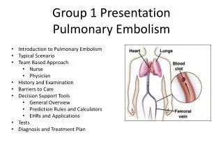
Pulmonary Embolism Presentation to Diagnosis. Objectives. Review the incidence, symptoms and presenting signs of PE Learn about clinical prediction models Learn about different diagnostic methods and diagnostic algorithms. Incidence.

Pulmonary Embolism Presentation to Diagnosis
E N D
Presentation Transcript
Pulmonary Embolism Presentation to Diagnosis
Objectives • Review the incidence, symptoms and presenting signs of PE • Learn about clinical prediction models • Learn about different diagnostic methods and diagnostic algorithms.
Incidence • The true incidence of PE is unknown and is suspected to be underestimated • It is estimated to be between 0.5% to 3% in the general population • Mortality from PE is estimated to be 0.1%
Risk Factors • Previous or current DVT • Immobilization • Surgery within the last 3 months • Stroke/paralysis • Central venous instrumentation within the last 3 months • Malignancy • CHF • Autoimmune diseases • Air travel * • Thrombophillias • In Women • Obesity (BMI ≥29) • Pregnancy • Heavy cigarette smoking (>25 cigarettes per day) • Hypertension
Presentation Most Common Symptoms Most Common Signs • Dyspnea at rest or with exertion (73 %) • Pleuritic pain (44 %) • Cough (34 %) • >2-pillow orthopnea (28 %) • Calf or thigh pain (44 %) • Calf or thigh swelling (41 %), • Wheezing (21 %) • Rapid onset of dyspnea • within seconds (46 %) • within minutes (26 %) • Tachypnea (54 %) • Tachycardia (24 %) • Rales (18 %), • Decreased breath sounds (17 %), • Accentuated pulmonic component of the second heart sound (15 %) • Jugular venous distension (14 %)
Case • A 63-year-old woman with stage IV lymphoma calls 911 for acute shortness of breath (SOB). At baseline, the patient is mobile and does not have SOB. She is also taking hormone replacement therapy. On the day of admission, she develops a sudden SOB and new pleuritic chest pain. She does not improve with nebulizer treatment on the way to the hospital. In the ER, her pulse is 115 bpm, RR = 36/min, temp = 100.1oF and O2 sat = 88% on room air. On exam, her lungs are clear, and her extremities are normal. A chest x-ray (CXR) shows mild right-sided atelectasis. An ABG shows ph = 7.48, PCO2 = 32 mm Hg and PO2 = 50 mm Hg on room air. • What is this patient’s pretest probability for having a pulmonary embolism? • What diagnostic method would you use to confirm this?
Clinical Decision Rules • Models for assessing clinical Probability of Pulmonary Embolism • Well’s Criteria • Geneva Score
D-Dimer • Elevated in thrombosis, malignancy, pregnancy, elderly, hospitalized patients • Role in low or moderate probability for PE • Normal results can rule out PE • Estimated 3 month risk of thromboembolism with negative D-dimer is 0.14% • Role in high probability patients proceed to CT, negative d-dimer can miss up to 15% of patients in this group
EKG in Pulmonary Embolism • Most commonly sinus tachycardia, with possible nonspecific ST/T wave changes • Only 10% of patients can have the S1Q3T3 so not reliable • Other EKG abnormalities including atrial arrhythmias, right bundle branch block, inferior Q-waves, and precordial T-wave inversion and ST-segment changes, are associated with a poor prognosis.
Chest Radiography • Not a sensitive or specific test for the diagnosis of PE. • Atelectasis, Pleural effusion, or a pulmonary parenchymal abnormality is noted most commonly • Only a small portion of patients with PE have a normal CXR.
Ventilation-Perfusion Scans • Useful if Normal (negative predictive value of 97%) • Also useful if High probability (positive predictive value of 85 to 90%) • Unfortunately, only diagnostic in 30 to 50% of patients
CT Angiography • Studies have shown sensitivity of close to 95% with an experienced observer • One of the most commonly cited benefits of CTA is its ability to detect alternative pulmonary abnormalities that may explain the patient's symptoms and signs • In 67% of patients without PE, CT provided additional information for alternate diagnosis • May predispose patients to further unnecessary testing
Pulmonary Angiography in PE • The “gold standard” • A negative pulmonary angiogram excludes clinically relevant PE. • The risk of embolization in patients with a negative angiogram is extremely low
Is it important to use clinical decision rules? • In the setting of no thromboembolic risk factors, it is extraordinarily unlikely (0.95% chance) to have a CT angiogram positive for PE. • With the combination of a negative D-dimer test result, this risk is even lower.
Lower Extremity US indicated? • Depends on pre-test probability • High pretest probablity for PE and negative CT may require additional testing • Good initial test to evaluate for pulmonary embolism in patients with contrast allergy, renal insufficiency, pregnancy, or critically ill patients. • Inexpensive test without radiation exposure • Can avoid additional testing if positive
Case Presentation • Reminder: • A 63-year-old woman with stage IV lymphoma with acute shortness of breath (SOB) and pleuritic chest pain. At baseline, the patient is mobile and does not have SOB. She is also taking hormone replacement therapy. In the ER, her pulse is 115 bpm. On exam, her lungs are clear, and her extremities are normal. A chest x-ray (CXR) shows mild right-sided atelectasis. An ABG shows ph = 7.48, PCO2 = 32 mm Hg and PO2 = 50 mm Hg on room air.
Case Presentation • Applying the Wells’ Scoring system, the patient has a moderate likelihood of having a PE with a score of 5.5 for high clinical suspicion for PE, tachycardia, and cancer. • Considering the patient’s score is >4, may proceed to CT angiography for PE rule out. • This patient did have CTA performed, which confirmed presence of PE. • She was subsequently started on anticoagulation
Summary and Recommendations • Consider your patient’s risk factors for pulmonary embolism • The clinical presentation of acute pulmonary embolism is variable and nonspecific • The major diagnostic tests employed in the evaluation of a patient with suspected PE include d-dimer testing, CTPA, V/Q scanning, venous ultrasonography, and conventional pulmonary angiography • Follow a diagnostic algorithm that combines CTPA, d-dimer and clinical assessment