Download
1 / 60
610 likes | 862 Vues
Circulatory System. Explain the structure of the heart. Analyze the function of the heart. Analyze circulation and the blood vessels. Discuss characteristics and treatment of common cardiac and circulatory disorders. bell. List functions of circulatory system. Bell….

E N D
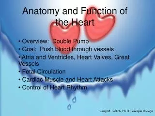
Circulatory System Explain the structure of the heart. Analyze the function of the heart. Analyze circulation and the blood vessels. Discuss characteristics and treatment of common cardiac and circulatory disorders.
bell • List functions of circulatory system
Bell…. • List the layers of the heart • Name the chambers
Functions • Pump • Blood transport system around body • Carries O2 and nutrients to cells, carries away waste products • Lymph system – returns excess tissue fluid to general circulation
BLOOD AND THE HEART FUN FACTS • An average adult human contains about 5 liters (5.3qt) of blood. • The blood makes up about one-thirteenth of the body’s weight. • The adult heart weighs about 280 grams (10 oz.) • At rest, the heart pumps out about 80 millimeters (2.6 oz) of blood with each beat. • The heart beats, on average, 70 times each minute at rest. • This means all the blood is circulated (goes round the body once) in about one minute. • During strenuous exercise the heart can pump six to eight times the amount of blood that it pumps at rest.
bell • List the three layers of the heart
The Heart • The Heart • Muscular organ • Size of a closed fist • Weighs 12-13 oz • Location – thoracic cavity • APEX – conical tip, lies on diaphragm, points left • Stethoscope – instrument used to hear the heartbeat
Structure • Hollow, muscular, double pump that circulates blood • At rest = 2 oz blood with each beat, 5 qts./min., 75 gallons per hour • Ave = 72 beats per minute • 100,000 beats per day
Bell • List all structures leading away from the heart…write whether the vessel is oxygenated (red) or deoxygenated (blue)
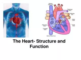
Structures Leading to and Away from….. • Superior vena cava and inferior vena cava – bring deoxygenated blood to right atrium • Pulmonary artery – takes blood away from right ventricle to the lungs for O2 • Pulmonary veins – bring oxygenated blood from lungs to left atrium • Aorta – takes blood away from left ventricle to rest of the body
Chambers • SEPTUM divides into R and L halves • Upper chambers – RIGHT ATRIUM and LEFT ATRIUM • Lower chambers – RIGHT VENTRICLE and LEFT VENTRICLE • Four heart valves permit flow of blood in one direction
The valves of the heart: superior view with the atria removed
Cross section of the heartValves-by order of flow TCV,PSV,BV,ASV
Blood Flow Through The Heart Veins/Venules SVC/IVC Right Atrium Tricuspid Valve Right Ventricle Pulmonic Valve Pulmonary Artery Lungs CO2 and O2 Exchange Pulmonary Veins Left Atrium Bicuspid Valve Left Ventricle Aortic Valve Aorta Arteries/Arterioles Capillaries to Body Blue Red F R O M L U N G S F R O M BO D Y TO LUNGS TO BODY
A. pericardium, B. myocardium, C. endocardium, D. septum, E. superior vena cava, F. inferior vena cava, G. right atrium, H. tricuspid valve, I. right ventricle, J. pulmonary valve, K. pulmonary arteries, L. pulmonary veins, M. left atrium, N. mitral or bicuspid valve, O. left ventricle, P. aortic valve, Q. aorta
Have A Heart • Label the:++++++++++++++++++++++++++ • septum • Superior and Inferior vena cava right atrium tricuspid valve right ventricle Pulmonary simi-lunar valve pulmonary artery • Oxygenated blood flows from lungs via pulmonary veins left atrium (bicusbid) mitral valve left ventricle Aortic similunar valve aorta
Payday/Bell ringer • Clean off your write on wipe off heart • Label all layers • Vessels • Valves • Chambers
Make a Heart 101 Your assignment is to work in you assigned groups (do not change them) and using the materials in your heart packet, construct a heart. You may use the finger tips of the gloves for your valves. You may use up to four cups, but you do not have to use all of these if you are doing something else creatively. Your heart must have all the chambers, valves, arteries, veins, etc. The note cards should be cut into small pieces and used as labels. Your heart must be labeled. The tape needs to be returned to the teacher at the end of class to be used with other classes. To complete this assignment you must write a group essay in which you use your particular model to teach a person about the heart. Make sure you make reference to the different structures and the materials you have used to make those structures. This essay will be graded for content, grammar, and spelling. Please use paragraphs. Use other people in your group to proof-read your essay. You have only this class period to complete this assignment, so do not waste time. For maximum success, put your heart into this assignment!
PHYSIOLOGY OF THE HEART • The heart is a double pump. When the heart beats… • Right Heart • Deoxygenated blood flows into heart from vena cava right atrium tricuspid valve right ventricle pulmonary semilunar valve pulmonary artery lungs (for oxygen) • Left Heart • Oxygenated blood flows from lungs via pulmonary veins left atrium mitral valve left ventricle aortic semilunar valve aorta general circulation (to deliver oxygen) • See page 287 • Systemic…Aortic, Arteries, Arterioles. Capillaries, Veinules. Veins, Superior and Inferior Vena Cava
Measuring and Recording PulseThe pressure of blood pushing against the wall of an artery as the heart beats and rests. Radial – base of thumb Temporal – side of forehead Carotid – side of neck Brachial – inner aspect of elbow Femoral – inner aspect of upper thigh Popliteal-behind knee Pedal-top of foot [Fill in E (3rd page)]
Heart Sounds = lubb dupp Heart Sounds = lubb dupp The sound you hear with your stethoscope is the sound the valves make as they open and close. • Apical Pulse • Taken with a stethoscope at the apex of the heart • Actual heartbeat heard and counted • Tips of earpieces and diaphragm of stethoscope should be cleaned with alcohol before use • Heart sounds heard resemble “lubb-dupp”
Bell • Put in order/sequence….the blood vessels after they have left the aorta, and before they go into the Superior and Inferior Vena Cava • 1) • 2) • 3) Capillaries • 4) • 5)
Circulatory system: heart,(Aorta) arteries, Arterioles, capillaries, Venules Veins (Sup/Inf Vena Cava)see page 267Capillary is Endothelial tissue
Venous circulationVeins-”low pressure”- vessels are thinner, more distended by pressure on them. Without pressure they collapse.
...use this slide to fill in outline 2nd pg A. 1-2 • CARDIOPULMONARY CIRCULATION -carries blood from heart to lungs • 1. Oxygenated and deoxygenated blood • 2. Oxygen/carbon dioxide exchange • Page 286
General/Systemic Circulation • 1. SYSTEMIC CIRCULATION – from the heart to the tissues and cells, then back to the heart • ARTERIOLES – small arteries • VENULES – small veins • Systemic Circulation PAGE 287 • AORTA – largest artery in the body • First branch is coronary artery • Aortic arch • Many arteries branch off the descending aorta • Where does it go? Circulation Wheel!
Using your wheel and information from your book, pg 262 answer (workbook) B.
General/Systemic Circulation ...use this slide to fill in outline 2nd pg C 1-5 • ARTERIES • Carry oxygenated blood away from the heart to the capillaries • Elastic, muscular and thick-walled • Transport blood under very high pressure • ARTERIOLES – small arteries • VEINS • Carry deoxygenated blood away from capillaries to the heart • Veins contain a muscular layer, but less elastic and muscular than arteries • Thin walled veins collapse easily when not filled with blood • Superior and Inferior vena cava carry blood to the heart • VENULES – small veins • CAPILLARIES • Smallest blood vessels, can only be seen with a microscope • Connect arterioles with venules PAGE 287 • Walls are one-cell thick and extremely thin – allow for selective permeability of nutrients, oxygen, CO2 and metabolic wastes • -------------------------------------------------------- • VALVES – permit flow of blood only in direction of the heart • JUGULAR vein – located in the neck
Circulation Race·Before the race starts, appoint a team captain. Have the team captain distribute the note cards, with the term facing down so the student does not know what it is. Make sure each team member has at least one term. Some may have more depending on the number of students per team. ·When the whistle blows, the students are to race to see who can create a “circulation circle” which follows the flow of blood through the circulatory system. Students can also race against their own times to see if they pick up speed.
Where does it go? • AORTA – largest artery in the body • First branch is coronary artery • Aortic arch • Many arteries branch off the descending aorta • Where does it go? Circulation Wheel!
Bell…”control of Heart contraction” • Read Conduction System…page 267 • List the 4 electrical impulses needed for a heart to beat
Your Heart’s Electrical System • http://www.nhlbi.nih.gov/health/dci/Diseases/hhw/hhw_electrical.html
Control of Heart Contractions • Page 267 text • SA (sinoatrial) NODE = PACEMAKER • Located in right atrium • SA node sends out electrical impulse • Impulse spreads over atria, making them contract • Travels to AV Node • AV (atrioventricular) NODE • Conducting cell group between atria and ventricle • Carries impulse to bundle of His • BUNDLE OF HIS • Conducting fibers in septum • Divides into R and L branches to network of branches in ventricles (Purkinje fibers) • PURKINJE FIBERS • Impulse shoots along Purkinje fibers causing ventricles to contract
ELECTROCARDIOGRAM (EKG or ECG) • Device used to record the electrical activity of the heart. • SYSTOLE = contraction phase • (Atrial Depolarization) • DIASTOLE = relaxation phase • (Ventricular relaxation) • Baseline of EKG is flat line • See page 248
Blood Pressure • Surge of blood when heart pumps creates pressure against the walls of the arteries • SYSTOLIC PRESSURE – measured during the contraction phase • DIASTOLIC PRESSURE – measured when the ventricles are relaxed • Average systolic = 120 • Average diastolic = 80 • PULSE – alternating expansion and contraction of an artery as blood flows through it. • Pulse sites: • BRACHIAL • CAROTID • RADIAL • POPLITEAL • PEDAL
Expert Groups • Topics: • 1)Rhythmic And Conduction (pg 253) • 2)CAD (pg 250) • 3)Infectious (pg 250) • 4)Blood Vessel (pg 277) • 5)Heart Failure (pg 252) • All same/like numbered get together and become experts (10 min) • Return original group. Share.Each person should have a page of notes, covering all topics.
Bell…. • Put your name on your work from Friday, pass up…a) Circulatory Disease Fact Chart • B) Pkt items H and I • C) Get out your outline…find diseases
Bell • Complete a worksheet called “The Heart”
ARRHYTHMIA (or dysrrhythmia) – any change from normal heart rate or rhythm BRADYCARDIA – slow heart rate (<60 bpm) TACHYCARDIA – rapid heart rate (>100 bpm) Diseases of the Heart
Coronary Artery Disease • ANGINA PECTORIS – chest pain, caused by lack of oxygen to heart muscle, treat with nitroglycerin to dilate coronary arteries • MYOCARDIAL INFARCTION • MI or heart attack • Lack of blood supply to myocardium causes damage • Due to blockage of coronary artery or blood clot atherosclerosis – plaque build-up on arterial walls, or arteriosclerosis – loss of elasticity and thickening of wall. • Amount of damage depends on size of area deprived of oxygen • Symptoms – severe chest pain radiating to left
Symptoms – severe chest pain radiating to left shoulder, arm, neck and jaw. Also nausea, diaphoresis, dyspnea. • Immediate medical care is critical • Rx – bedrest, oxygen, medication • Morphine for pain, tPA to dissolve clot • Anticoagulant therapy to prevent further clots from forming • Angioplasty and by-pass surgery may be necessary • Friday, Feb. 4, 2010 is National Wear Red Day - Everyone (Men too - Think about your mother, wife, daughter, sister, aunt, etc) can support the fight against heart disease in women by wearing red on Friday. Heart Disease is the No. 1 killer of American women. For more information check out the website http://goredforwomen.org/
Treatment • Heart Surgery • CORONARY BY-PASS – usually, a healthy vein from the leg removed and attached before and after the coronary obstruction, creating an alternate route for blood supply to the myocardium.
Balloon angioplasty is used to reopen a blocked coronary artery • Coronary arteries supply blood to the heart tissue
An artificial pacemaker is implanted under the skin • PACEMAKERS • Demand pacemaker – fires only when heart rate drops below programmed minimum • CPR – cardiopulmonary resuscitation, used in the presence of cardiac arrest