Download
1 / 25
260 likes | 468 Vues
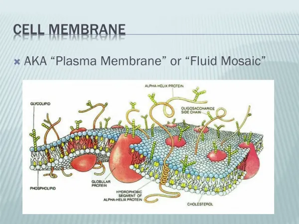
Development of a Device to Measure Cell Membrane Water Permeability. BY ROBERT ELDER MENTOR: DR. ADAM HIGGINS. Permeability. Permeability is a property of the cell membrane Measures how easily a substance can cross the membrane Different for each substance and cell type

E N D
Development of a Device to Measure Cell MembraneWater Permeability BY ROBERT ELDER MENTOR: DR. ADAM HIGGINS
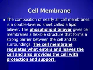
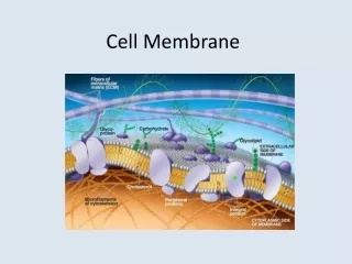
Permeability • Permeability is a property of the cell membrane • Measures how easily a substance can cross the membrane • Different for each substance and cell type • The cell membrane is relatively permeable to water • Significance of water permeability: • Cryopreservation • Biosensing
Significance for Cryopreservation • Cryopreservation: techniques that can keep biological matter intact for years • Water permeability affects the amount of water in a cell, which affects viability • Too much water → intracellular ice → physical damage • Too little water → high concentration → chemical damage • Cryoprotectants that alter permeability are a possibility • Current cryogenic techniques are successful only with cell suspensions, where isolated cells float in fluid
Significance for Biosensors • Certain toxins form membrane pores that increase permeability • Examples: • Plague bacterium • Staphylococcus aureus • A device to detect permeability changes could be used to detect these toxins
Measuring Permeability • The Coulter principle • Particles flowing through a channel change the electrical resistance in proportion to their size - + Resistance momentarily increased Ω Conductive solvent Suspended cell flows through channel
Measuring Permeability • Concentration differences across membranes cause water to flow, which causes cells to shrink or expand • Isotonic – same concentration inside and outside cell results in no net water flow
Measuring Permeability • The rate of the volume change is related to the membrane permeability (P) • P is permeability • C is total solute concentration • Z is a collection of other constants
Measuring Permeability Overview • Goal: build a device to measure permeability by using the Coulter principle to determine volume changes. • Resistance → Volume → Permeability Isotonic solution Hypotonic solution Electrical current
Existing Measurement Methods • Examples • Fluorescence quenching • Concentration change of marker molecules due to cell uptake • Problems • Complex modifications to sample • Specialized equipment • Not portable • Time consuming • A more convenient method should be developed to accelerate research efforts
Project Goals • Complete device construction and characterization • Develop a model to relate resistance changes to permeability • Compare our measurements to those from established techniques (fluorescence quenching) • Test the effect of different substances on permeability
Device Overview • Dual inlet system for switching solutions quickly • Heat exchanger to control temperatures • Channel: 100µm deep for sensitive measurements Electrodes + - Flow Channel Syringes Heat Exchanger Cell Monolayer
Device Design Heat exchanger shell Clear plastic allows microscopy Coverslip and gasket form flow channel
Device Characterization: Solution Exchange • How long does it take to switch solutions? • Relevance • Cells respond to concentration changes in seconds • Switching concentrations must be much faster • Method • Use dye solutions to visualize solution exchange • Compare to mathematical model of diffusion and fluid flow
Device Characterization: Solution Exchange • Model results • Solution exchange is much faster than volume changes • Dye exchange results • Slower than model results • Solution exchange less than 1 second at relevant flowrates Channel Top Chamber Entrance Point of Interest Channel Bottom
Device Characterization: Heat Exchanger • Dual inlet system can result in rapid temperature changes in channel • Initially, isotonic solution pumped: temperature depends on heat exchanger • Then, anisotonic solution pumped: temperature equal to shell temperature Initial: Flowing Syringes Heat Exchanger Not flowing
Device Characterization: Heat Exchanger • Dual inlet system can result in rapid temperature changes in channel • Initially, isotonic solution pumped: temperature depends on heat exchanger • Then, anisotonic solution pumped: temperature equal to shell temperature Final: Not Flowing Syringes Heat Exchanger Flowing
Device Characterization: Heat Exchanger • Goal: minimize the initial temperature change when switching solutions (i.e. get isotonic temperature equal to anisotonic) • Method: increase tube length, investigate tube material Goal: Flowing Syringes Heat Exchanger Not flowing
Device Characterization: Heat Exchanger Long Medium Short
Fluorescence Quenching Measurements • Purpose: obtain permeability measurements for comparison • Fluorescence intensity is directly related to cell volume • Fluorescence is quenched (decreased) when hypertonic solution shrinks cells
Fluorescence Quenching Measurements • Relative intensity changes can be used to determine permeability Intensity Time (s)
Fluorescence Quenching Measurements • Relative intensity changes can be used to determine permeability Normalized Intensity Time (s) Time (s)
Effect of Cytochalasin D on Permeability • A cell-permeable mycotoxin • Potent inhibitor of actin polymerization • Changes cell morphology and possibly permeability • Possible cryoprotectant but tests inconclusive
Results • Response of resistance measurements was too slow to measure volume changes accurately • Further work may be pursued to decrease response time • Fluorescence quenching experiments were successful and gave results of the predicted order of magnitude
Acknowledgements • Howard Hughes Medical Institute • University Honors College • Dr. Adam Higgins • Dr. Kevin Ahern • Nick Lowery, Crystal Gupta, Logan Williams • Andy Brickman, Manfred Dittrich