Animal Development
Animal Development. The question of how a zygote becomes an animal has been asked for centuries As recently as the 18th century, the prevailing theory was called preformation

Animal Development
E N D
Presentation Transcript
The question of how a zygote becomes an animal has been asked for centuries • As recently as the 18th century, the prevailing theory was called preformation • Preformation is the idea that the egg or sperm contains a miniature infant, or “homunculus,” which becomes larger during development
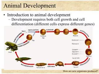
WHAT DETERMINES DEVELOPMENT • Development is determined by the zygote’s genome and differences between embryonic cells • Cell differentiation is the specialization of cells in structure and function • Morphogenesis is the process by which an animal takes shape
Big ideas • Gametes (fertilizaiton) • Zygote (cleavage) • Blastula (gastrulation) • Gastrula (neurulation) • Organogenesis • Role of genes & protein concentration gradients • Induction: communication from an inducer to a competent responder
Fertilization • 2 major events: • Fertilization brings the haploid nuclei of sperm and egg together, forming a diploid zygote • The sperm’s contact with the egg’s surface initiates metabolic reactions in the egg that trigger the onset of embryonic development • Most info comes from sea urchin studies • External fertilization • Problems of external fertilization: • Dilution/protection of gametes in the enormous volume of the ocean • Correct species fertilization • Blocking polyspermy
The Acrosomal Reaction • The acrosomal reaction is triggered when the sperm meets the egg • This reaction releases hydrolytic enzymes that digest material surrounding the egg • Acrosomal process adheres to receptors on vitelline layer (species specific) • Sperm/egg membranes fuse, sperm nucleus enters • Na+ influx, depolarization • Depolarization sets up fast block to polyspermy
Contact and fusion of sperm and egg membranes Entry of sperm nucleus Acrosomal reaction Sperm plasma membrane Fast block polyspermy Sperm nucleus Cortical reaction Contact Acrosomal process Basal body (centriole) Sperm head Fertilization envelope Fused plasma membranes Cortical granule Actin Perivitelline space Hydrolytic enzymes Acrosome Jelly coat Cortical granule membrane Vitelline layer Sperm-binding receptors Egg plasma membrane EGG CYTOPLASM
The Cortical Reaction • Fusion of egg and sperm also initiates the cortical reaction • This reaction induces a rise in Ca2+ in cytoplasm that stimulates cortical granules to release their contents outside the egg • Corticalgranules fuse w/ membrane • Enzymes • Polysaccharides • Fertilization envelope formed = slow block to polyspermy (follows repolarization) • These changes cause formation of a fertilization envelope that functions as a slow block to polyspermy
500 µm Fast block polyspermy 30 sec 10 sec after fertilization 20 sec 1 sec before fertilization Spreading wave of calcium ions Point of sperm entry
Activation of the Egg • The sharp rise in Ca2+ in the egg’s cytosol increases the rates of cellular respiration and protein synthesis by the egg cell • Chemical signals from cortical rxn cause H+ to be transported out --> increase in pH • Nuclei fuse • Egg/sperm differences • Egg contains proteins, mRNA not found in sperm • Ca2+ injection, temperature shock can cause artificial activation • With these rapid changes in metabolism, the egg is said to be activated
1 Binding of sperm to egg Acrosomal reaction: plasma membrane depolarization (fast block to polyspermy) 2 3 4 6 Seconds 8 10 Increased intracellular calcium level LE 47-5 20 Cortical reaction begins (slow block to polyspermy) 30 40 50 Formation of fertilization envelope complete 1 2 Increased intracellular pH 3 4 Increased protein synthesis 5 Minutes 10 20 Fusion of egg and sperm nuclei complete 30 Onset of DNA synthesis 40 60 First cell division 90
Fertilization in Mammals • In mammalian fertilization, the cortical reaction modifies the zona pellucida as a slow block to polyspermy
LE 47-6 Follicle cell Sperm basal body Cortical ganules Zona pellucida Sperm nucleus Egg plasma membrane Acrosomal vesicle EGG CYTOPLASM
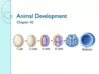
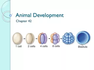
Cleavage • Fertilization is followed by cleavage, a period of rapid cell division without growth • Cleavage partitions the cytoplasm of one large cell into many smaller cells called blastomeres
LE 47-7 Morula Fertilized egg Blastula Four-cell stage
The eggs and zygotes of many animals, except mammals, have a definite polarity • The polarity is defined by distribution of yolk, with the vegetal pole having the most yolk • The development of body axes in frogs is influenced by the egg’s polarity
Animal hemisphere Animal pole Point of sperm entry Vegetal hemisphere Vegetal pole LE 47-8 Point of sperm entry Future dorsal side of tadpole Anterior Gray crescent Right First cleavage Ventral Dorsal Left Posterior Body axes Establishing the axes
Cleavage planes usually follow a pattern that is relative to the zygote’s animal and vegetal poles
Zygote 0.25 mm 2-cell stage forming LE 47-9 4-cell stage forming Eight-cell stage (viewed from the animal pole) 8-cell stage 0.25 mm Animal pole Blasto- coel Blastula (cross section) Vegetal pole Blastula (at least 128 cells)
Meroblastic cleavage, incomplete division of the egg, occurs in species with yolk-rich eggs, such as reptiles and birds
Disk of cytoplasm Fertilized egg Zygote Four-cell stage LE 47-10 Blastoderm Cutaway view of the blastoderm Blastocoel BLASTODERM YOLK MASS Epiblast Hypoblast
Holoblastic cleavage, complete division of the egg, occurs in species whose eggs have little or moderate amounts of yolk, such as sea urchins and frogs
Gastrulation • Gastrulation rearranges the cells of a blastula into a three-layered embryo, called a gastrula, which has a primitive gut
The three layers produced by gastrulation are called embryonic germ layers • The ectoderm forms the outer layer • The endoderm lines the digestive tract • The mesoderm partly fills the space between the endoderm and ectoderm Video: Sea Urchin Embryonic Development
The mechanics of gastrulation in a frog are more complicated than in a sea urchin-INVAGINATION • OTHERS- INVOLUTION
CROSS SECTION SURFACE VIEW Animal pole Blastocoel Dorsal tip of blastopore Dorsal lip of blastopore Vegetal pole Blastula LE 47-12 Blastocoel shrinking Archenteron Ectoderm Mesoderm Blastocoel remnant Endoderm Key Future ectoderm Future mesoderm Yolk plug Yolk plug Gastrula Future endoderm
Gastrulation in the chick and frog is similar, with cells moving from the embryo’s surface to an interior location • During gastrulation, some epiblast cells move toward the blastoderm’s midline and then detach and move inward toward the yolk. INVOLUTION
Epiblast LE 47-13 Primitive streak Future ectoderm Endoderm Migrating cells (mesoderm) Hypoblast YOLK
Organogenesis • During organogenesis, various regions of the germ layers develop into rudimentary organs organs
Early in vertebrate organogenesis, the notochord forms from mesoderm, and the neural plate forms from ectoderm Video: Frog Embryo Development
Neural folds LE 47-14a LM 1 mm Neural fold Neural plate Notochord Ectoderm Mesoderm Endoderm Archenteron Neural plate formation
Neural plate Neural fold The neural plate soon curves inward, forming the neural tube LE 47-14b Neural crest Outer layer of ectoderm Neural crest Neural tube Formation of the neural tube
Somites Eye Tail bud • Mesoderm lateral to the notochord forms blocks called somites • Lateral to the somites, the mesoderm splits to form the coelom SEM 1 mm LE 47-14c Neural tube Notochord Neural crest Coelom Somite Archenteron (digestive cavity) Somites
Eye Neural tube Notochord Forebrain LE 47-15 Somite Heart Coelom Archenteron Endoderm Lateral fold Mesoderm Blood vessels Ectoderm Somites Yolk stalk YOLK Yolk sac Form extraembryonic membranes Neural tube Early organogenesis Late organogenesis
Many structures are derived from the three embryonic germ layers during organogenesis
Developmental Adaptations of Amniotes • Embryos of birds, other reptiles, and mammals develop in a fluid-filled sac in a shell or the uterus • Organisms with these adaptations are called amniotes • In these organisms, the three germ layers also give rise to the four membranes that surround the embryo
Amnion Allantois Embryo Amniotic cavity with amniotic fluid Albumen LE 47-17 Shell Yolk (nutrients) Yolk sac Chorion
Mammalian Development • The eggs of placental mammals • Are small and store few nutrients • Exhibit holoblastic cleavage • Show no obvious polarity • Gastrulation and organogenesis resemble the processes in birds and other reptiles • Early cleavage is relatively slow in humans and other mammals
At completion of cleavage, the blastocyst forms • The trophoblast, the outer epithelium of the blastocyst, initiates implantation in the uterus, and the blastocyst forms a flat disk of cells • As implantation is completed, gastrulation begins • The extraembryonic membranes begin to form • By the end of gastrulation, the embryonic germ layers have formed-ECTODERM, MESODERM AND ENDODERM
Endometrium (uterine lining) Inner cell mass Trophoblast Blastocoel Blastocyst reaches uterus. LE 47-18a Expanding region of trophoblast Maternal blood vessel Epiblast Hypoblast Trophoblast Blastocyst implants.
Expanding region of trophoblast Amniotic cavity Amnion Epiblast Hypoblast Chorion (from trophoblast Yolk sac (from hypoblast) LE 47-18b Extraembryonic membranes start to form and gastrulation begins. Extraembryonic mesoderm cells (from epiblast) Allantois Amnion Chorion Ectoderm Mesoderm Endoderm Yolk sac Extraembryonic mesoderm Gastrulation has produced a three-layered embryo with four extraembryonic membranes.