Optimizing FSC Thresholds for Murine Lymphocyte and Myeloid Cell Analysis by Flow Cytometry
This study outlines the method for optimizing forward scatter (FSC) thresholds to accurately identify murine lymphocytes and myeloid cells using the LSR-II flow cytometer. Post-sample collection, FlowJo software was utilized to gate out aggregates using FSC-A vs. FSC-H plots. Cells were then gated based on CD45 positivity and viability dye exclusion to ensure only live cells were analyzed. Specific cell populations, such as neutrophils, were further assessed by selecting cells negative for TCRβ and CD19/B220 while evaluating surface markers like CD11b and Gr1.

Optimizing FSC Thresholds for Murine Lymphocyte and Myeloid Cell Analysis by Flow Cytometry
E N D
Presentation Transcript
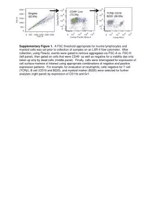
Supplementary Figure 1. A FSC threshold appropriate for murine lymphocytes and myeloid cells was set prior to collection of samples on an LSR-II flow cytometer. After collection, using FlowJo, events were gated to remove aggregates via FSC-A vs. FSC-H (left panel), then gated on cells that were CD45+ as well as negative for a viability dye only taken up only by dead cells (middle panel). Finally, cells were interrogated for expression of cell surface markers of interest using appropriate combinations of negative and positive expression patterns. For example, for evaluation of neutrophils, cells negative for T cell (TCRb), B cell (CD19 and B220), and myeloid marker (B220) were selected for further analyses (right panel) by expression of CD11b and Gr1.