Image manipulation
Image manipulation. Definition. Those techniques (operations) or process which modify an image or group of images to enhance the visibility of useful information while suppressing (noise) or non-useful information. Image manipulation.

Image manipulation
E N D
Presentation Transcript
Definition Those techniques (operations) or process which modify an image or group of images to enhance the visibility of useful information while suppressing (noise) or non-useful information.
Image manipulation Do not produce any additional information. The information content in the processed image is always less than or equal to that in the original image.
Image manipulation techniques: Two categories: -Linear----Such as image smoothing and enhancement. -Non-linear----grey scale manipulation (to be modified by different algorithms)
Non-linear techniques: The algorithm is based on a point processing technique referred to a grey level mappingwhich is referred to with different names but windowing is the most common image processing technique in CT
Windowing A method by which CT image grey scale can be manipulated using the CT numbers of the image. The operator can alter these numbers to provide an optimum demonstration of the different structures.
Windowing: The picture contrast is easily changed with two control mechanisms: The window width and window level.
Window width (WW): The absorption measurements range in CT (HU). It determines the maximal number of shades of grey that can be displayed on the CT monitor.
Window level (WL): The centre or midpoint of the range of CT numbers and can be positioned any where on the WW.
Large window width: Indicates that there is a relatively long grey scale or a large block of CT numbers that will be assigned value of grey.
Narrow window width: Implies that the transition from black to white will take place over a relatively few CT numbers.
Manipulating WW and WL -WW is 2000 -1000 CT numbers above 0 and another 1000 CT numbers below 0 -The mid point of the range (WL) is 0, it is referred to as a reference point.
CT numbers range varies between scanners: -1000 to +3095 HU (4095 CT numbers) -2048 to +6143 HU (8191 CT umbers)
-The grey scale changes as the WW is expanded or narrowed: -Bone structures WW include higher CT numbers on the scale. -Structures that contain air, WW must include lower CT numbers on the scale.
Effect of WW on image contrast: • As the WW increases the contrast decreases • As the WW decreases the contrast become greater • Contrast is optimized with medium WW settings.
Effects of WL on image display: • As the WL increases the picture changes from white to black. • As the WL moves towards the higher CT numbers (white), more CT numbers with lower values (black) are displayed.
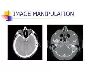
WW and WL manipulation WW 2000 WL 300 WW 90 WL 35
Lung detail No Lung detail
Low contrast window High contrast window Bone window
Radiation dose: Radiation dose must be known to estimate the patients potential risk from radiation and to weigh this risk against the benefits of CT scanning.
Radiation dose Radiation dose from CT scanner examination are among the highest in diagnostic radiology.
Dose measurement methods: The ionization chamber method is the easiest and probably the most accurate and it is used almost exclusively to report dose
Ionization chamber Consists of a small air-filled container with thin walls that allow radiation to pass through easily. As the high-energy photons (x-rays) collide with air molecules enclosed within the ionization chamber, some molecules are “ionized” (electrons are knocked from some molecules)
Ionization chamber These free electrons can be collected on a conducting wire or plate and measured as electric charge. The amount of collected charge is proportional to the amount of ionization, which is proportional to the amount of radiation that pas through the chamber.
Ionization chamber The charge is removed from the ionization chamber and measured with a very sensitive instrument known as an electrometer It is measured in coulombs.
Multiple Scan Average Dose (MSAD) A series of CT scans are performed on patient, between each scan the patient is moved a bed index (BI) distance. Each slice delivers the characteristic bell-shaped dose curves. If the dose from all scans are summed, the resulting total patient dose resembles the oscillating curve.