
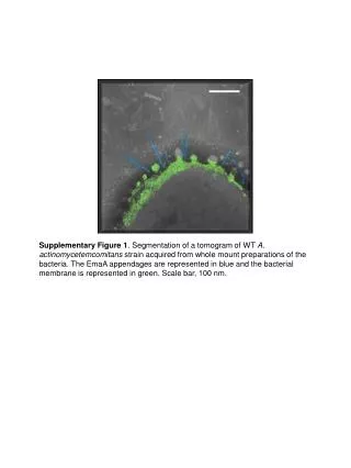
Supplementary Figure 1 . Segmentation of a tomogram of WT A. actinomycetemcomitans strain acquired from whole mount preparations of the bacteria. The EmaA appendages are represented in blue and the bacterial membrane is represented in green. Scale bar, 100 nm.

E N D
Presentation Transcript
Supplementary Figure 1. Segmentation of a tomogram of WT A. actinomycetemcomitans strain acquired from whole mount preparations of the bacteria. The EmaA appendages are represented in blue and the bacterial membrane is represented in green. Scale bar, 100 nm.
Supplementary Figure 2. Electron micrographs of A. actinomycetemcomitans strains acquired from whole mount preparations of the bacteria stained with 2% phosphotungstic acid (pH 7). A: WT strain (wild type strain); B: the emaA mutant strain transformed with pKMΔ502-534; C: the emaA mutant strain transformed with pKMΔ535-610; D: the emaA mutant strain transformed with pKMΔ611-641. Arrows point to the apical end of the EmaA appendages. Inserts, windowed EmaA appendages. Spherical particles in the images are extracellular vesicles. BM, bacterial membrane. Scale bar, 100 nm.
Supplementary Figure 3. EmaA appendages windowed from electron micrographs of A. actinomycetemcomitans strains acquired from whole mount preparations of the bacteria stained with 2% phosphotungstic acid (pH 7). A: WT strain (wild type strain); B: emaA mutant strain transformed with pKMΔ70-206 (plasmid containing an in-frame deletion corresponding to amino acids 70-206). Bracket indicates the long head domain, including Sub-domain I and Sub-domain II. Arrows points to a broadening at the distal end of the stalk domain. BM, bacterial membrane. Scale bar, 10 nm.
Supplementary Figure 4. Domain prediction of EmaA obtained by submitting the sequence to the daTAA server (http://toolkit.tuebingen.mpg.de/dataa), a Trimeric Autotransporter Adhesin domain prediction program. AutoSP: autotransporter signal peptide; Yhead: forms a beta-roll; Neck: connector domain that joins beta structures with coiled-coils; HIN2: Head FxG motif; Trp ring: transition between coiled-coils and beta meanders.