Download
1 / 34
350 likes | 1.65k Vues
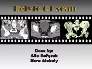
The Pelvic Scan: Early pregnancy problems Gynaecological abnormalities. Normal pelvic anatomy. Anatomy of uterus and ovaries Arterial blood supply Venous blood supply Neural supply. Gynaecological scan indications. Pelvic pain Pelvic mass Irregular/ heavy vaginal bleeding

E N D
The Pelvic Scan:Early pregnancy problemsGynaecological abnormalities
Normal pelvic anatomy • Anatomy of uterus and ovaries • Arterial blood supply • Venous blood supply • Neural supply
Gynaecological scan indications • Pelvic pain • Pelvic mass • Irregular/ heavy vaginal bleeding • Post menopausal vaginal bleeding • Infertility • Endocrine symptoms/ signs • Recurrent miscarriage
Routine • Sagittal image uterus and bladder • Length of endometrium and cervix • Measure length uterus +/- ET in AP • Check for fluid in POD • Transverse image bladder and cervix • Transverse image bladder and uterus at widest part • Image whole uterus as move cephalad • Measure ET in AP • Measure uterus width and AP • Transverse to top of uterus
Routine • Transverse image broad ligament • Move to Right/Left • Check no tubal dilatation • Visualise ovary • Move caudally if unable to see • Split screen and measure ovarian volume • Cyst? • Measure volume • Septations, papillary lesions, solid areas, low level internal echoes • Check mobility with valsalva or hand on abdomen • Check for ascites • Torsion?
Early pregnancy scan indications • Dating • Bleeding • Pain • Previous ectopic/ miscarriage/ molar pregnancy
Complete miscarriage Bleeding and cramps which are usually settling
Missed miscarriage Spotting only usually. Expected to be 6-12 weeks by LMP. Fetal pole seen
Other miscarriages • Anembryonic pregnancy • Spotting or nil • Gestational sac, MSD >2cm • No fetal pole • Incomplete miscarriage • Bleeding and cramps • RPOC • Doppler to diagnose • Threatened miscarriage • Bleeding +/- pain • Viable pregnancy
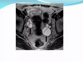
Ectopic pregnancy • Pregnancy outside the uterine corpus • Ampulla • Cornu • Ovary • Abdominal • Life- threatening intra-abdominal bleeding • Symptoms and signs • Spotting, pain- usually one side, fainting, shouler-tip pain • URGENT referral on USS diagnosis • High index of suspicion if • previous ectopic • IUD • infertility
USS findings • Empty uterus • Adnexal mass • +/- FHR • Ring of blood flow on doppler • Tenderness on probe pressure over mass • Free fluid especially POD • TV scan ideally if available
Hydatidiform molar pregnancy • Abnormal placental development • Usually no recognisable fetus • ‘Snowstorm’ appearance on USS • Exaggerated symptoms of pregnancy • Hyperemesis • Thyroid hormone abnormality • Large theca-lutein cysts • Rx is ERPOC and CXR • Can recur and rarely in malignant form