X-ray Scattering from Thin Films
470 likes | 1.86k Vues
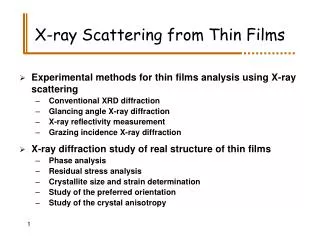
X-ray Scattering from Thin Films. Experimental methods for thin films analysis using X-ray scattering Conventional XRD diffraction Glancing angle X-ray diffraction X-ray reflectivity measurement Grazing incidence X-ray diffraction X-ray diffraction study of real structure of thin films

X-ray Scattering from Thin Films
E N D
Presentation Transcript
X-ray Scattering from Thin Films • Experimental methods for thin films analysis using X-ray scattering • Conventional XRD diffraction • Glancing angle X-ray diffraction • X-ray reflectivity measurement • Grazing incidence X-ray diffraction • X-ray diffraction study of real structure of thin films • Phase analysis • Residual stress analysis • Crystallite size and strain determination • Study of the preferred orientation • Study of the crystal anisotropy
Conventional X-ray diffraction Diffracting crystallites • + Reliable information on • the preferred orientation of crystallites • the crystallite size and lattice strain (in one direction) • - No information on the residual stress (constant direction of the diffraction vector) • - Low scattering from the layer (large penetration depth)
Glancing angle X-ray diffraction GAXRD Gold, CuKa, m 4000 cm-1 Symmetrical mode GAXRD
Other diffraction techniques used in the thin film analysis Conventional diffraction with W-scanning qy=0 Grazing incidence X-ray diffraction (GIXRD) qz»0 Conventional diffraction with C-scanning qx=0
Penetration depth of X-rays L.G. Parratt, Surface Studies of Solids by Total Reflection of X-rays, Physical Review 95 (1954) 359-369. Example: Gold (CuKa) d = 4.2558 10-5 b = 4.5875 10-6
r t [Å]s [Å] 0.68 19.6 5.8 0.93 236.5 34.0 1.09 14.1 2.7 1.00 5.0 2.7 1.00 2.8 X-ray reflectivity measurement Calculation of the electron density, thickness and interface roughness for each particular layer Mo Edge of TER Mo Kiessig oscillations (fringes) Mo W Si The surface must be smooth (mirror-like)
Angle of incidence, g Sample Goebel mirror Sample inclination, y X-ray source Diffraction angle, 2q Sample rotation, f Diffraction vector Scintillation detector Normal direction Flat monochromator Experimental set-up Used for XRR, SAXS, GAXRD and symmetrical XRD
Information on the microstructure of thin films • Study of the anisotropy in the lattice deformation • Investigation of the depth gradients of microstructure parameters • Phase analysis • Residual stress analysis • Crystallite size and strain determination • Study of the preferred orientation
Uranium nitride – phase analysis Sample deposition PVD in reactive atmosphere N2 Heated quartz substrate (300°C) • Phase composition • UN, 80-90 mol.% • Fm3m, a = 4.8897 Å • U2N3, 10-20% mol.% • Ia3, a = 10.64 - 10.68 Å Schematic phase diagram 800 T(°C) 400 UN2 U2N3 U UN 0 Atomic Percent Nitrogen 50 60 67
U2N3 versus UN2 U2N3 (Ia3), a = 10.66 Å U: 8b (¼, ¼, ¼) U: 24d (-0.018, 0, ¼) N: 48e (0.38, 1/6, 0.398) UN2 (Fm3m) a = 5.31 Å U: 4a (0, 0, 0) N: 8c (¼, ¼, ¼) Cannot be distinguished in thin films U N
Uranium nitride – residual stress analysis U2N3 • a0 = (10.636 ± 0.002) Å • Compressive residual stress • s = - (6.2 ± 0.1) GPa • No anisotropy of lattice deformation UN • a0 = (4.926 ± 0.015) Å • Compressive residual stress • s = - (1.8 ± 0.8) GPa • Strong anisotropy of lattice deformation GAXRD at g=3°
Uranium nitride – anisotropic lattice deformation UN • a0 = (4.9270 ± 0.0015) Å • s = - (1.0 ± 0.1) GPa 111 easy hard directions
UN – anisotropic lattice deformation Dependence of the lattice deformation on the crystallographic direction R.W. Vook and F. Witt, J. Appl. Phys., 36 (1965) 2169. Related to the anisotropy of the elastic constants
UN versus U2N3 Anisotropy of the mechanical properties is related to the crystal structure UN (Fm3m) a = 4.93 Å U: 4a (0, 0, 0) N: 4b (½, ½, ½) U2N3 (Ia3), a = 10.66 Å U: 8b (¼, ¼, ¼) U: 24d (-0.018, 0, ¼) N: 48e (0.38, 1/6, 0.398) U N
Methods for the size-strain analysis using XRD Lattice strain • Local changes in the d-spacing • Line broadening increases with increasing q (a result of the Bragg equation in the differential form) Crystallite size • Fourier transformation of finite objects (with limited size) • Constant line broadening (with increasing diffraction vector) (011) (011) (111) (111) (001) (001) (110) (110) (000) (100) (100) (000) Scherrer Williamson-Hall Warren-Averbach Krivoglaz P. Klimanek (Freiberg) R. Kuzel (Prague) P. Scardi (Trento) T. Ungar (Budapest)
UN – anisotropic line broadening • The Williamson-Hall plot • It recognises the anisotropy of the line broadening • It is robust (weak intensity, overlap of diffraction lines) • It is convenient if the higher-order lines are not available (nanocrystalline thin films, very thin films, GAXRD) 100 111
UN – texture measurement Reciprocal space mapping Preferred orientation {110}
9 3 3 3 8 {111} 4 2 2 7 5 1 1 3 3 1 6 4 2 0 2 2 2 5 5 -1 1 3 3 -1 3 1 1 [1/A] z q 4 4 -2 2 4 0 0 2 2 0 3 5 -1 -1 3 -1 1 1 1 1 2 4 -2 0 -2 2 2 2 0 0 -3 3 1 3 -1 -1 -1 1 1 1 4 -2 -2 -2 2 0 0 -8 -7 -6 -5 -4 -3 -2 -1 0 1 q [1/A] x Reciprocal space mapping A highly textured gold layer Measured using CuKa radiation
Epitaxial growth of SrTiO3 on Al2O3 SrTiO3: Fm3m 111 axis -3 001Al2O3: R-3c Reciprocal space map Atomic ordering in direct space Sr O in SrTiO3 Ti O in Al2O3 Al
SrTiO3 on Al2O3 Atomic Force Microscopy Pyramidal crystallites with two different in-plane orientations 111 111 _ 110 _ 110 AFM micrograph courtesy of Dr. J. Lindner, Aixtron AG, Aachen.
TiN TiC TiN WC Depth resolved X-ray diffraction TiN TiC TiCN Absorption of radiation
Surface modification of thin films Gradient of the residual stress in thin TiN coatings (CVD) implanted by metal ions: Y, Mo, W, Al and Cr
Functionally graded materials W. Lengauer and K. Dreyer, J. Alloys Comp. 338 (2002) 194 Nitrogen – in-diffusion from N2 N-rich zone of (Ti,W)(C,N) Ti(C,N) N-poor zone of (Ti,W)(C,N) (Ti,W)C SEM micrograph courtesy of C. Kral, Vienna University of Technology, Austria
Study of concentration profiles The lattice parameter must depend on concentration Copper radiation Penetration depth: 1.8 mm Molybdenum radiation Penetration depth: 12.5 mm
SummaryBenefits of X-ray scattering ... for investigation of the real structure of thin films • Length scale between 10-2Å and 103Å is accessible (from atomic resolution to the layer thickness) • Small and variable penetration depth of X-ray into the solids (surface diffraction, study of the depth gradients) • Easy preparation of samples, non-destructive testing • Integral measurement (over the whole irradiated area)