Download

1 / 19
2.08k likes | 10.98k Vues
PTERYGIUM. PREPARED BY : FARHUDA ZULAIKHA BT DOL @ ABDUL WAHID DURATUL’AIN BT MOHD NAZRI NURUL NAJUWA BT SAZALI. PTERYGIUM. DEFINITION ETIOLOGY PATHOLOGY CLINICAL PICTURE (Signs & Symptoms, course) DIFFERENTIAL DIAGNOSIS TREATMENT. 1) DEFINITION.

E N D
PTERYGIUM PREPARED BY : • FARHUDA ZULAIKHA BT DOL @ ABDUL WAHID • DURATUL’AIN BT MOHD NAZRI • NURUL NAJUWA BT SAZALI
PTERYGIUM • DEFINITION • ETIOLOGY • PATHOLOGY • CLINICAL PICTURE (Signs & Symptoms, course) • DIFFERENTIAL DIAGNOSIS • TREATMENT
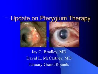
1) DEFINITION • Most often refers to a benign growth of conjunctiva • It grows from the nasal side of sclera • pterygium is a fleshy growth that invades the cornea (the clear front window of the eye). It is an abnormal process in which the conjunctiva (a membrane that covers the white of the eye) grows into the cornea.
2) ETIOLOGY • Unknown but may related to : a) Ultraviolet rays (most important factor) -engaging in occupations that require outdoor activity b) Chronic irritation by dust etc c) Genetic predisposition -development of pterygium appear to exist in certain families
3) PATHOLOGY • Pterygium consists of: - Degeneration of collagen -Fibro-vascular proliferation (fibrous tissue + blood vessels) -overlying covering of epithelium (thin layer)
4) CLINICAL PICTURE a- Signs & Symptoms b- Course
APEX : blunt and lies over the cornea and may grow to reach papillary area(affects vision) Signs • Site : On the nasal side less commonly temporal and always bilateral • Shape : Is triangular and consists of apex, neck & base BASE : Lies over the sclera NECK : Overlies the limbus
Course A) PROGRESSIVE B) STATIONARY • Thick and fleshy • Vascular • Preceded by white dots • Thin and membranous • Less vascular
5) DIFFERENTIAL DIAGNOSIS A- Neoplasia # carcinoma in situ of conjunctiva and cornea. Also known as Bowen’s disease. # squamouscell carcinoma # other neoplasticdiseases B- Pannus C- Pingueculae # commonly occurring, generally small and asymptomatic # occasionally be subject to some inflammation with symptoms of itching, burning, or mild pain # mild-to-moderate focal thickening of the conjunctivalstroma with elastotic degeneration of collagen
pterygium pingueculae
6) TREATMENT • Pterygium is best left alone - if small & stationary - recurrence may follow the operation • Indications of operations : # progressive # if it encroaches pupillary area # if patient is annoyed (cosmetic reason)
NON SURGICAL • 1) Eyedrops for irritation and redness. • 2) Protection from sunlight and dust to prevent it from wosening • 3) Steroids to lessebn inflammations
Operation for pterygium • Excision + bare sclera technique • pterygium is removed leaving a raw area of sclera • (this allows corneal epithelium to cover the cornea before conjunctival epithelium reaches the limbus) • Excision + application mucous membrane,conjunctival or coeneal graft • - gap in the mucous membrane (conjunctiva) tissue, where the pterygium was removed, is filled with a transplant of tissue that has been painlessly removed from underneath the upper eyelid.
PREVENTION OF PTERYGIUM • Wearing protective sunglasses with side shields and/or wide brimmed hats (as it is associated with excessive sun or wind exposure) • Using artificial tears (prevent their formation/stop further growth • Surfers & water-sport athletes: - wear eye protection (block 100% of UVR from water)
MEASURES USED TO PREVENT RECURRENCE : • Expose the bare area to Beta irradiation. • Expose the bare area to Mytocin C • Excision & mucous membrane grat. • Excixion & lamellar (structural) keratoplasty • Stem cell transplantation