Skull: Anterior View
Skull: Anterior View. Figure 7.2a. Skull: Posterior View. Figure 7.2b. Parietal Bones and Major Associated Sutures. Form most of the superior and lateral aspects of the skull. Figure 7.3a. Occipital Bone and Its Major Markings. Forms most of skull’s posterior wall and base
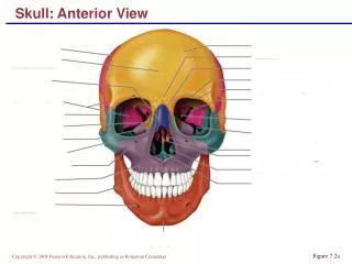

Skull: Anterior View
E N D
Presentation Transcript
Skull: Anterior View Figure 7.2a
Skull: Posterior View Figure 7.2b
Parietal Bones and Major Associated Sutures • Form most of the superior and lateral aspects of the skull Figure 7.3a
Occipital Bone and Its Major Markings • Forms most of skull’s posterior wall and base • Major markings include the posterior cranial fossa, foramen magnum, occipital condyles, and the hypoglossal canal Figure 7.2b
Occipital Bone and Its Major Markings Figure 7.4b
Temporal Bones Figure 7.5
Sphenoid Bone Figure 7.6a, b
Ethmoid Bone Figure 7.7
Mandible and Its Markings Figure 7.8a
Maxillary Bone Figure 7.8b
Anterior Aspects of the Skull Figure 7.2a
Posterior Aspects of the Skull Figure 7.2b
External Lateral Aspects of the Skull Figure 7.3a
Midsagittal Lateral Aspects of the Skull Figure 7.3b
Inferior Portion of the Skull Figure 7.4a
Inferior Portion of the Skull Figure 7.4b
Orbits Figure 7.9b
Nasal Cavity Figure 7.10a
Nasal Cavity Figure 7.10b
Paranasal Sinuses Figure 7.11
Vertebral Column Figure 7.13
Vertebral Column: Ligaments Figure 7.14a
Vertebral Column: Intervertebral Discs Figure 7.14b
General Structure of Vertebrae Figure 7.15
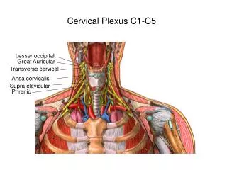
Cervical Vertebrae Table 7.2
Cervical Vertebrae: The Atlas (C1) Figure 7.16a, b
Cervical Vertebrae: The Axis (C2) Figure 7.16c
Cervical Vertebrae: The Atlas (C2) Figure 7.17a
Thoracic Vertebrae Figure 7.17b
Lumbar Vertebrae Figure 7.17c
Sacrum and Coccyx: Anterior View Figure 7.18a
Sacrum and Coccyx: Posterior View Figure 7.18b
Bony Thorax (Thoracic Cage) Figure 7.19a
Bony Thorax (Thoracic Cage) Figure 7.19b
Cervical Vertebrae Table 7.2
Cervical Vertebrae: The Atlas (C1) Figure 7.16a, b
Cervical Vertebrae: The Axis (C2) Figure 7.16c
Cervical Vertebrae: The Atlas (C2) Figure 7.17a
Thoracic Vertebrae Figure 7.17b
Lumbar Vertebrae Figure 7.17c
Sacrum and Coccyx: Anterior View Figure 7.18a
Sacrum and Coccyx: Posterior View Figure 7.18b
Bony Thorax (Thoracic Cage) Figure 7.19a
Structure of a Typical True Rib • Bowed, flat bone consisting of a head, neck, tubercle, and shaft Figure 7.20
Pectoral Girdles (Shoulder Girdles) Figure 7.22a
Clavicles (Collarbones) Figure 7.22b, c
Scapulae (Shoulder Blades) Figure 7.22d, e
Humerus of the Arm Figure 7.23
Bones of the Forearm Figure 7.24
Radius and Ulna Figure 7.24