Download
1 / 32
320 likes | 455 Vues
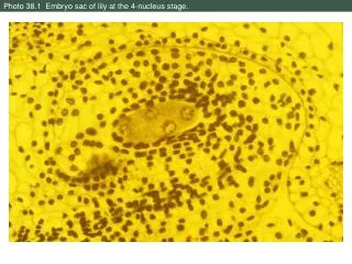
Photo 38.1 Embryo sac of lily at the 4-nucleus stage. Photo 38.2 Mature embryo sac of lily. Photo 38.3 Cross-section through anther of monocot, showing pollen. LM. Photo 38.4 Cross section through anther of a eudicot, showing pollen. LM.

E N D
Photo 38.3 Cross-section through anther of monocot, showing pollen. LM.
Photo 38.4 Cross section through anther of a eudicot, showing pollen. LM.
Photo 38.5 Pores and furrows in highly magnified grass pollen. SEM.
Photo 38.6 Pollen of passion flower (Passiflora racemosa) stained with fluorescent dye.
Photo 38.7 Flowering inflorescence of a grass; anthers exerted for wind dispersal of pollen.
Photo 38.8 Stigma of Hibiscus sp.; pollen grains adhere to sticky stigma. SEM.
Photo 38.10 Pollen tube germinated in vitro on artificial medium. LM, Nomarski.
Photo 38.11 Endosperm of a kernel of corn; starch darkly stained. LM.
Photo 38.12 Development of shepherd’s purse (Capsella bursa-pastoris). LM.
Photo 38.17 Fruits of hawkweed (Hieracium floribundum), family Asteraceae. SEM.
Photo 38.18 Sprouting coconuts on a beach on Rangiroa Atoll, Tahiti.
Photo 38.19 Fruit of enchanter’s nightshade (Circaea quadrisulcata). SEM.
Photo 38.20 Chrysanthemum sp. buds form more readily under short-day conditions than long-day
Photo 38.21 Vegetative reproduction: stolons of spider plant with new plants at tip.
Photo 38.22 Vegetative reproduction: daffodil bulb. White storage leaves, yellow flower buds.
Photo 38.24 Vegetative reproduction: Plantlets form at edge of leaf of Kalanchoe daigremontiana.
Photo 38.25 Vegetative reproduction: grass rhizomes in Goolwa Sand Dunes, Australia.
Photo 38.26 Dandelion heads, with asexually produced seeds ready for dissemination on the wind.
Photo 38.27 Each clump of aspen is a single modular organism.
Photo 38.28 Amaryllis belladonna has a perfect flower, with clearly visible carpel and stamens.
Photo 38.29 Male (left) and female (right) flowers of zucchini, an imperfect monoecious plant.
Photo 38.30 Male flower of the pussy willow (Salix capraea), an imperfect dioecious plant.
Photo 38.31 Female flower of the pussy willow (Salix capraea), an imperfect dioecious plant.
Photo 38.32 Cross section through kernels of corn, Zea mays, showing embryo and endosperm.