Download
1 / 97
1.15k likes | 2.47k Vues
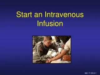
Chapter 13 Intravenous Infusion and Blood Transfusion. SECTION ONE Intravenous Infusion. Definition:IV infusion is a method that a large volume of solution is infused into vein to correct fluid and electrolyte disturbance . solution ; passage (infusion set); vein. Intravenous Infusion.

E N D
SECTION ONEIntravenous Infusion • Definition:IV infusion is a method that a large volume of solution is infused into vein to correct fluid and electrolyte disturbance. solution; passage(infusion set); vein
Intravenous Infusion • IV infusion is a serious and complex responsibility that requires the nurse: • proficiency in performance • familiarity with the anatomy involved • mindful use of principles of asepsis • expertise in prevention, management of complications that may occur with treatment
Intravenous Infusion • Fluids are medications--IV infusion requires a physician’s order • the type and amount of fluid administered will be based on types of patient’s need the patient’s age general health status the results of laboratory tests
Types of solutions There are many methods of classification. • according to their osmolality in relation to normal blood plasma • according to their purpose Hypotonic fluids Isotonic fluids Hypertonic fluids Nutrient solutions Electrolyte solutions Volume Expanders
Hypotonic fluids • have lower osmolality than plasma • to correct dehydration as they move from blood vessels into the cells • examples are 0.45 % NaCl, 0.2 % NaCl, or 5%GS • excessive infusion can cause water intoxication
Isotonic fluids • have the same effective osmolality as plasma • to expand the intravascular space to correct hypovolemia as in shock • examples are lactated Ringer’s, 0.9 % ( normal ) saline(0.9%NaCl), 5 % dextrose in normal saline(5%GNS). 1.4%NaHCO3 • excessive infusion can cause circulatory overload and pulmonary edema
Hypertonic fluids • have greater osmolality than plasma • to pull fluid from cells and the interstitial space into the intravascular space to relieve edema • examples are >5 % dextrose solutions, colloidal products such as dextran, 3 % saline ( rarely used ). • excessive infusion can cause cellular dehydration and circulatory overload or diuresis.
Nutrient solutions • contain some form of glucose and water for calories and fluids replacement • examples are 5%or 10%dextrose in water Hypertonic ( >10 percent dextrose ) parenteral nutrition solutions are irritating to peripheral veins and so must be infused into central veins.
Electrolytes solutions • contain varied amounts of cations and anions • examples are normal saline,Ringer’s solution, and lactated Ringer’s solution • commonly be used to restore vascular vo1ume,particularly after trauma or surgery • also be used to replace fluid and electrolytes for patients with continuing losses,for example,gastric suction or wound drainage
Volume expanders • be used to increase the blood volume following severe loss of blood or loss of plasma • examples aredextran,plasma,and human serum albumin
Clinical routine • In clinic, prepare fluids fall into the following three categories: • Crystalloid Solution • Colloidal Solution • Parenteral Nutrition Solutions
Crystalloid Solution • have small molecular weights and stay in blood vessel for a short time • maintain the balance of fluids in intracellular and extra- cellular • correct the fluids and electrolytes disturbance • commonly used crystalloid solutions are: • Dextrose in Water Solutions • Isotonic Electrolytes Solutions • Alkaline solutions • Hypertonic Solutions
Dextrose in Water Solutions • Be used for fluids and calories replacement,decreasing the consumption of albumen,and preventing the production of ketone. • glucose is decomposed quickly in body, usually doesn’t cause hypertonic and diuretic effects. • Clinically there are usually 5%GS and 10%GS (25%GS; 50%GS rarely used)
Isotonic Electrolytes Solutions • be used in electrolytes replacement. • Loss of body fluids usually is accompanied with disturbance of electrolytes.So, the balance of fluids and electrolytes must be maintained during fluids replacement. • examples are o.9%NaCl,Ringer’s isotonic solution and 5% GNS.
Alkaline solutions • NaHC03 in Water Solutions • Be used in correcting acidosis, and regulating of acid-base balance. • NaHC03 Na+ and HCO3- HCO3- + H+ H2CO3 C02 +H2 O. • Commonly used solutions are 5%NaHC03 and 1.4% NaHC03 Solutions. • Sodium Lactate in Water Solutions • The concentrations of the solution usually used in clinic are 11.2%and 1.84%.
Hypertonic Solutions • be used for diuretic and dehydration purposes • increase osmolality of blood plasm pulling fluids into plasma reduce the edema of tissues • can decrease intracerebral pressure and improve the function of central nervous system. • Clinically,mannitol 20%,sorbitol 25%and dextrose 25%-50%in water solutions are often used.
Colloidal Solution • have large molecular weight,can stay in blood for a long time • can maintain plasma colloid osmotic pressure effectively, • expand the blood volume, • improve microcirculation, • elevate the blood pressure. • examples are :dextran plasma substitutes blood products
Dextran • It is water-soluble polysaccharide of high molecular polymer. • moderate molecular dextran : elevate plasma colloid osmotic pressure,expand blood volume • small molecular dextran : reduce the viscosity of blood,decrease the accumulation of erythrocytes,improve microcirculation and tissue perfusion volume,and prevent the formation of thrombosis
Plasma Substitutes • can expand vascular volume and cardiac output greatly • can be used with whole blood in acute massive hemorrhage • examples are 706 (hydroxyethylamylum),povidone and oxypolygelation
Blood Products • can elevate colloid osmotic pressure • expand vascular volume • provide protein and antibody • help with tissue repair and enhance immunity of body
Parenteral Nutrition Solutions • be intravenously given to the patients who are unable to get nutrition via gastrointestinal tract or have inadequate intake of nutrients • provide calories,proteins,vitamins and minerals,and maintain the balance of nitrogen • main compositions : amino acids,fatty acids,vitamins,minerals,high concentration of glucose ,and water • commonly used solutions : multiple amino acids solutions , fat emulsions
sequence principle of solution transfusion • First crystalloid solutions then colloidal solutions • First sodium chloride solutions then dextrose in water solutions • first fast then slow, rather shortage than overload • rather acid than alkaline • Potassium solutions properly
Sites of Venipuncture • Peripheral Superficial Vein • veins in dorsal hands : the first choice for adult patients • median cubital,basilic,cephalic veins :drawing blood bolus injections of medication PICC • The saphena veins in legs and veins in dorsal feet are not the first choice because of the danger of thrombosis caused by the vein valve. • Veins in dorsal foot are commonly used for children,but are avoided in adults because of the danger of thrombophlebitis. • Veins in the Scalp :for infants • Subclavian,External Jugular:for central venous access
peripheral intravenous infusion • Purposes • Preparation nurse patient environment equipment • Procedures and Key Points • EvaluationVCD
Purposes • To correct or prevent fluid and electrolyte disturbances resulted from illnesses, altered fluid intake, or prolonged episodes of vomiting or diarrhea. • To increase the blood volume, maintain blood pressure following severe loss of blood, severe burns, or shock. • To supply medication to cure diseases for rapid effectiveness. • To supply nutrient substances to promote wound healing, weight gain and positive nitrogen balance for patients with chronic consuming illness, inability to intake, digest or absorb a diet. • To establish a lifeline for rapidly needed medications.
Preparation • Nurse 1. Review the physician’s order in patient’s record. 2.Evaluate patient’s age and medical status. Evaluate patient’s renal status and other pertinent lab data (e.g., electrolyte, serum glucose ). 3. Wash hands and wear mask. • patient 1. Verify patient’s identity. 2. Explain the procedure and purpose.Ask the patient to void. 3. Position the patient for comfort and optimal visibility for skill performance.
Preparation • Environment:cleanness, commodiousness and brightness • equipment: Medical tray ·Antiseptic solution ·Sterile swab ·Tourniquet ·infusionPad ·Adhesive tape ·File and vial opener ·IV solution and medication ·Bottle bag ·Infusion set ·kidney-shaped tray
Procedures and Key Points 1.Check and right • the bed number,the patient’s name,medication name, concentration, dosage, dateand usage • the quality of solution(the cap of bottle, the expiration date , deposition,cloudiness, foreign matter, any cranny on the bottle’s body) 2. Complete the medication label and stick it to the solution container
3. Add medications into solution 4.Insert the infusion set 5. Prepare the equipment and take them to the bedside. Check again 6. Discharge air
7.Select the venipuncture site(pad , tourniquet ) 8.Sterilize the venipuncture site.Prepare adhesive tape. 9.Check again. 10. venipuncture The wizened The obese The elderly The dropsical (edema)
11.fixation 12. Regulate the flow rate 13. Check again. 14. Disposure after operation (equipment,patient) 15. Change bottles 16. Disposure after infusion (equipment,patient)
Cautions 1. Follow the principles of asepsis and check system strictly to prevent infection and mistakes. 2. Arrange the sequence of IV fluids rationally according to the patient’s need. Assign medications according to the therapeutic principles and the half life of medications. 3. Protect and use veins reasonably (usually from small veins) to patients who need long-term IV infusion.
Cautions 4. Prevent air embolism by ejecting air thoroughly in infusion set, changing fluid bottles and withdrawing the venipuncture needle in time. 5. Assess for compatibility of medications. Ensure the needle have been inserted into vein before administration irritative or special medications. 6. Master the flow rate strictly. 7. Assess during infusion carefully in order to find the problems and settle the problems on time. Document the result after assessment.
Education 1. Tell the patient don’t regulate the flow rate optionally. 2. Introduce the signs and symptoms of complications with IV reactions, ask patient call nurse in time when he find the signs of IV reaction. 3. Instruct the patient to report any blood in the tube,a stoppage in the flow,or increased discomfort. 4. Intensify mental nursing to patient who need long-term IV infusion.
Evaluation • Assess the status of the skin and dressing of IV site by observing whether there is heat,pain,redness,or swelling • Whether the IV flows smoothly and whether the flow rate is consistent with what is ordered • Check the information to ensure the right medication administered • Signs and symptoms of complications with intravenous infusion • Patient’s knowledge about medication and infusion • Ability to perform self-care activities.
Regulating the Infusion Flow Rate • Calculate the Flow Rate • Common Infusion Control Device
Calculate the Flow Rate • Total time of infusion (h) = Total infusion vo1ume (m1)×drop factor (drops/m1) Drops per minute×60 • Drops per minute= Total infusion vo1ume (m1)×drop factor (drops/m1) Total time of infusion (min)
Calculate the Flow Rate Slow flow rate is suitable for • the elderly,infants and patients with diseases in heart, 1ungs,or kidney. • When hypertonic solutions,solutions containing potassium,or solutions containing medications for raising blood pressure are infused,the flow rate also should be slow. When a patient with normal heart and lung function has severe dehydration,the flow rate should be rapid.
Common Infusion Control Device • Clamp:be easy to operate; not precise • Infusion Pump: • exert positive pressure on the tubing or on the fluid to ensure measured amount of fluid is infused uniformly in a given time • has a drop sensor, and an alarm that will sound if drops are not detected at the appropriate rate VCD
Assessing during infusion The responsibilities of nurses in assessment are: • keeping the system sterile; • changing solution,tube,and dressings on time; • assisting the patient with self-care activities so as not to disrupt the system; • instruct the patient to report any blood in the tube,a stoppage in the flow,or increased discomfort.
Common Problems during Infusion and Methods to Treat • Slow Flow Rate or No Infusion • Too Large Volume of Solution in Chamber • Too Small Volume of Solution in Chamber • The Surface of Liquid Fall down Automatically assess the site and the infusion rate at least once an hour
Slow Flow Rate or No Infusion • Infiltration • occlusion of the IV Needle or Catheter • hyperkinesia of Vein • Too Low Hydrostatic Pressure
Infiltration • Cause:the needle dislodge from the vein and fluid exude in the subcutaneous space • Signs: insertion site becomes swollen,clammy, and painful • Alternative nursing actions: :discontinue IV and establish a new line at a new site
occlusion of the IV Needle • causes: • there are clots at the tip of the needle • the needle tip is against the vein wall (flexed joint-wrist , elbow ) • narrowing the tubing may exist too-tight IV dressing a kink in the tubing • measures: • assess : lowering the IV container below the level of the IV insertion site, opening the roller clamp thoroughly, and observing for a blood return • Alternative nursing actions: inspect the area around the insertion site loose the IV dressing check the tubing change the position of the needle handle or extremity
hyperkinesia of Vein • Causes: • extremity is exposed in cold environment for a long time • the temperature of the fluid is too low • Alternative nursing action: warm the extremity