Basics of Rietveld Refinement
791 likes | 2.78k Vues
Basics of Rietveld Refinement. Scott A Speakman 13-4009A x3-6887 speakman@mit.edu. Uses of the Rietveld Method.

Basics of Rietveld Refinement
E N D
Presentation Transcript
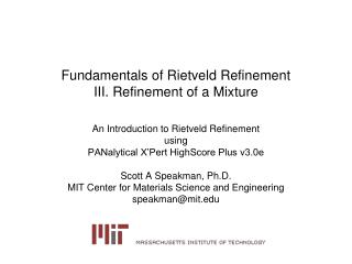
Basics of Rietveld Refinement Scott A Speakman 13-4009A x3-6887 speakman@mit.edu
Uses of the Rietveld Method • The Rietveld method refines user-selected parameters to minimize the difference between an experimental pattern (observed data) and a model based on the hypothesized crystal structure and instrumental parameters (calculated pattern) • can refine information about a single crystal structure • confirm/disprove a hypothetical crystal structure • refine lattice parameters • refine atomic positions, fractional occupancy, and thermal parameter • refine information about a single sample • preferred orientation • refine information about a multiphase sample • determine the relative amounts of each phase
Requirements of Rietveld Method • High quality experimental diffraction pattern • a structure model that makes physical and chemical sense • suitable peak and background functions
Obtaining High Quality Data • issues to consider • aligned and calibrated instrument • beam overflow problems • thin specimen error • good counting statistics • appropriate step size • sample transparency • surface roughness • preferred orientation • particle size • go to XRD Basics pg 102
Describing the Crystal Structure • space group • lattice parameters • atomic positions • atomic site occupancies • atomic thermal parameters • isotropic or anisotropic
The Crystal Structure of LaB6 • LaB6 • Space Group Pm-3m (221) • Lattice Parameter a=4.1527 A
Where to get crystal structure information • check if the structure is already solved • websites • Inorganic Crystal Structure Database (ICSD) http://icsd.ill.fr/icsd/index.html • 4% is available for free online as a demo • Crystallography Open Database http://www.crystallography.net/ • Mincryst http://database.iem.ac.ru/mincryst/index.php • American Mineralogist http://www.minsocam.org/MSA/Crystal_Database.html • WebMineral http://www.webmineral.com/ • databases • PDF4 from the ICDD • Linus Pauling File from ASM International • Cambridge Structure Database • literature • use the PDF to search ICSD listings and follow the references • look for similar, hopefully isostructural, materials • index the cell, and then try direct methods or ab-initio solutions • beyond the scope of today’s class
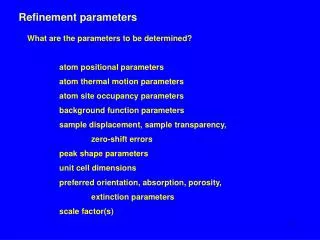
Instrumental Parameters • background • peak profile parameters • cagliotti parameters u, v, w • pseudo-voigt or other profile parameters • asymmetry correction • anisotropic broadening • error correcting parameters • zero shift • specimen displacement • absorption • extinction • roughness • porosity
How many parameters can we refine? • Each diffraction peak acts as an observation • theoretically, refine n-1 parameters • refining a tetragonal LaNi4.85Sn0.15 crystal structure, there might be: • scale factor • 2nd order polynomial background: 3 parameters • 2 lattice parameters • no atomic positions (all atoms are fixed) • 3 or 5 thermal parameters • 2 or 4 occupancy factors • zero shift and specimen displacement • 5 profile shape parameters • 22 parameters maximum with 43 peaks (20 to 120 deg 2theta) • does this mean we can refine all parameters?
background functions • manually fit background • polynomial • chebyshev • shifte chebyshev • amorphous sinc function • many others for different programs
profile functions • vary significantly with programs • almost all programs use Cagglioti U, V, and W • HSP uses pseudo-voigt, Pearson VII, Voigt, or pseudo-voigt 3 (FJC asymmetry) • GSAS uses functions derived more from neutron and synchrotron beamlines
How do you know if a fit is good? • difference pattern • Residuals R • R is the quantity that is minimized during least-squares or other fitting procedures • Rwp is weighted to emphasize intense peaks over background • Rexp estimates the best value R for a data set • an evaluation of how good the data are • RBragg tries to modify the R for a specific phase • GOF (aka X2)
Refinement Strategy • Rietveld methods fit a multivarialbe structure-background-profile model to experimental data • lots of potential for false minima, diverging solutions, etc • need to refine the most important variables first, then add more until an adequate solution is realized • a correct solution may not result …
Ray Young’s Refinement Strategy • scale factor • zero shift or specimen displacement (not both) • linear background • lattice parameters • more background • peak width, w • atom positions • preferred orientation • isotropic temperature factor B • u, v, and other profile parameters • anisotropic temperature factors
HSP Automatic Refinement Strategy • Very similar to Prof Young’s recommendations • a good choice for beginners • you can set limits on any of these parameters
Additional Files • XRD_Basics_HSP_2006.pdf • large collection of information about X-ray diffraction, instrumentation, and different techniques • X’Pert HighScore Plus Tutorial.pdf • overview of the different functionality available in HighScore Plus • Introduction.pdf • overview of Rietveld • parameters_calc_patterns.pdf • overview of parameters involved in calculating a diffraction pattern
further reading • “Rietveld refinement guidelines”, J. Appl.Cryst.32 (1999) 36-50 • R.A. Young (ed), The Rietveld Method, IUCr 1993 • V.K. Pecharsky and P.Y. Zavalij, Fundamentals of Powder Diffraction and Structural Characterization of Materials, Kluwer Academic 2003. • DL Bish and JE Post (eds), Modern Powder Diffraction, Reviews in Mineralogy vol 20, Min. Soc. Amer. 1989. • CCP14 website http://www.ccp14.ac.uk/tutorial/tutorial.htm • prism.mit.edu/xray/resources.htm
Rietveld Programs • Free • GSAS + ExpGUI • Fullprof • Rietica • PSSP (polymers) • Maud (not very good) • PowderCell (mostly for calculating patterns and transforming crystal structures, limited refinement) • Commercial • PANalytical HighScore Plus • Bruker TOPAS (also an academic) • MDI Jade or Ruby
Examples • Silicon • LaB6 • intermetallic LaNi4.85Sn0.15
Silicon • Open the datafile in HSP • Add the structure model • insert the structure manually • import (insert) a struture file • usually use the CIF format– the ubiquitous standard for crystal structures • HSP can also import ICSD *.cry files and structures from other refinement programs • GSAS can import CIF or PowderCell files • try the automatic refinement • manually improve the fit
Silicon Crystal Structure • Fd3m • which setting? (2) • a=5.43 A • Si at 0.125, 0.125, 0.125
Lanthanum hexaboride LaB6 • Open the datafile • insert the crystal structure CIF file • Note that boron (z=5) makes little difference in the XRD pattern compared to the lanthanum (z=57) • what can we do to improve the fit
LaNi4.85Sn0.15 • The data was taken from Chapter 6 of Fundamentals of Powder Diffraction and Structural Characterization of Materials, by Pecharsky and Zavalij • The structure is a bit more complex that our earlier example, which allows us to explore more features of HighScore Plus • The data (Ch6_1.raw) is in GSAS format, which can be read into HighScore Plus • I have also included a CIF file from the ICSD (#104685) with all the main features of the structure described
Issue to Consider • How can I work without knowledge of the structure? • Use LeBail or Pawley method to determine lattice parameters • Try indexing and solving the structure using the HighScore Plus tools • You will find that there are 16 possible space groups for this material, but picking the most common (and simplest) choice, P6/mmm, is the right way to go • Where do I put the atoms? • You can use a Fourier map to find out wherein the structure the electron densities are greatest. Put the heaviest atoms (La) at these sites, then work your way through the chemistry • What variables do I refine and in what sequence? • Take a look at the “automatic” option in HSP - this is not a bad strategy to use. We will go through these in detail…