Download
1 / 72
730 likes | 1.4k Vues
Total Cholesterol LDL-Cholesterol HDL-Cholesterol Lipoprotein(a) Homocysteine Particle size/density Lp subclasses Triglycerides Inflammatory factors. Oxidized-LDL (Ox-LDL) Metabolic syndrome Prothrombotic factors Apolipoproteins Glucose impairment Subclinical atherosclerotic diseases

E N D
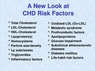
Total Cholesterol LDL-Cholesterol HDL-Cholesterol Lipoprotein(a) Homocysteine Particle size/density Lp subclasses Triglycerides Inflammatory factors Oxidized-LDL (Ox-LDL) Metabolic syndrome Prothrombotic factors Apolipoproteins Glucose impairment Subclinical atherosclerotic diseases Diabetes mellitus Life-habit risk factors A New Look at CHD Risk Factors
Measure LDL-C More Accurately Friedewald Calculation: • lLDL = TC – (HDL + VLDL) • (TG/5 approximates VLDL) • Recommendation: Not valid with TG > 400 mg/dL • l LDL = Lp(a) + IDL + Remnant Lp + LDL • lTotal Error = Bias + (1.96 x CVImprecision) • < 12% (ideal: + 4% & CV < 4%) • From: Clin Chem 41:1414-1420(1995)
Accuracy of LDL-c Calculation Method Triglycerides Percent Correct Percent Mg/dL (Within + 10%) Incorrect < 20093%7% 201-300 75% 25% 301-400 61% 39% 401-500 41% 59% > 500 < 20% > 80% From: Warnick GR et. al. Clin Chem 36:15-19 (1990)
Accuracy of LDL-c Calculation Method TriglyceridesCorrectLDL-C Incorrect LDL-C mg/dL (Within + 10%) < 200 85% 15% 201-300 77% 23% 301-400 59% 41% 401-600 41% 59% From: Warnick GR et. al. Clin Chem 36:15-19 (1990)
Friedewald Misclassifies Risk Category When Triglycerides > 177 mg/dL LDL Risk Category Marniemi J et al. Clin Biochem 1995 June; 28:285-289.
When LDL-C Target is < 100 Friedewald Should Not Be Utilized Directly Measured LDL Cholesterol 187 146 109 75 180 135 93 61 *Scharnagl H et al. Clin Chem Lab Med 2001 May;39(5):426-31.
Evaluation of Four Homogeneous Direct LDL-C Methods • Study: LDL-C methods from Genzyme, Reference Diagnostics (RD), Roche, and Sigma were evaluated for precision, accuracy, and specificity for LDL in the presence of abnormal lipoproteins. • Results: Precision was < 2% CV for all methods; and correlation to the CDC reference method was r = 0.955-1.067; total error was Genzyme = 12.6%, RD = 16.5%, Sigma = 38.3%, Roche = 41.6%. • Conclusion: The methods show nonspecificity toward abnormal lipoproteins, thus compromising accuracy. These direct methods are no better than the Friedewald LDL-C. • From:Miller, WG et.al. Clin Chem 48:489-498(2002)
Review of LDL-C Methods 1. Ultracentrifugation: Reference method, accuracy based Time-consuming, tedious, technically difficult, costly 2. Electrophoresis: Simultaneous separation of major lipoprotein fractions, can quantitate, visualize some unusual bands Technically difficult to do; unless automated, it can be tedious and time consuming, can be costly + - + -
Review of LDL-C Methods(Cont.) 3. Third Generation Direct Methods: No pretreatment, full automation, improved analytical precision, fasting specimen not required, save on labor cost Can have lack of LDL specificity; inaccuracy because of analytical interferences from TG, bilirubin, IDL-C, Lp-X, VLDL-C, Lp(a), apo E-rich HDL, and/or hemoglobin; can be costly From: Nauck M et. al. Clin Chem48:236-254 (2002) + -
HDL-c Testing • NCEP Laboratory Goals • Total Error = ≤ 13% (analytical bias +1.9% CV) • TE = Ideal: ≤ 5% and ≤ 4% CV • CDC Reference Method: 3 Stage Procedure • Ultracentrifuge = remove chylomicron + VLDL • Heparin MnCl2 ppt. = remove apoB lipoprotein • Abell-Kendall cholesterol assay on HDL • CRMLN Designated Comparison Method • Dextran Sulfate (50,000 DA) ppt. • Abell Kendall cholesterol assay on HDL
Homogenous HDL-c Methods • There are 4 types of homogeneous HDL-C methods: immunologic, PEG, synthetic polymer, enzymatic. • All can be automated & directly measure HDL. • Most have excellent precision (< 3% CV). • Many have CRMLN certification, suggesting that accuracy is possible with proper instrument, reagents, and calibrator. • Specificity and interferences: most are robust and can tolerate TG < 900 mg/dL, but biases can occur with atypical lipoprotein patterns. From: Warnick et. al. Clin Chem 47:1579-1596 (2001)
Review of HDL-c Methods 1. Ultracentrifugation: Comparison method for accuracy Time-consuming, tedious, technically difficult, costly, can have interference from Lp(a) and others 2. Electrophoresis: Simultaneous separation of major lipoprotein fractions, can quantitate, visualize some atypical bands Technically difficult to do; unless automated, it can be tedious and time consuming, can be costly + - + -
Review of HDL-C Methods(Cont.) 3. Third Generation Direct Methods: No pretreatment, full automation, improved analytical precision (<3% CV), fasting specimen not required, save on labor cost (20% savings) Can have lack of HDL specificity; inaccuracy because of analytical interferences from bilirubin, hemoglobin, high TG (chylomicrons and VLDL-c), Lp(a), apo E-rich HDL, and/or HDL variants (Apo A-IMilano) From: Nauck M et. al. Clin Chem48:236-254 (2002) + -
New Approaches to Quantitative Lipoprotein Electrophoresis • Completely automated; better precision • Better gel/buffer system (high-resolution) • Precision control of temperature during electrophoresis • Densitometer designed for higher sensitivity • Simultaneously measures VLDL-c, LDL-c, HDL-c & Lp(a)-c • LDL-c and HDL-c compares well with CDC reference methods • Lp(a) compares well with Lp(a) candidate reference method
Within-Run with Helena Electrophoretic Method Pool VLDL-C LDL-C HDL-C mg/dL mg/dL mg/dL B 8.1 ± 0.4 149.1 ± 2.0 41.4 ± 1.5 (5.3% CV) (1.3% CV) (3.6% CV) From: Naito HK, et al. Handbook of Lipoprotein Methods. AACC Press, Washington D.C., Pgs. 477-495(1997)
Comparison of Methods: Helena Electrophoresis vs. CDC Reference Method Fractions Regression r VLDL-C y=0734 + 0.389x 0.97682 LDL-C y=1.783 + 1.07x 0.99809 HDL-C y=2.449 + 1.1x 0.99241 From: Naito HK, et al. Handbook of Lipoprotein Methods. AACC Press, Washington D.C., Pgs. 477-495(1997)
Comparison of Methods: Helena vs. Ultracentrifugation Fractions Regression r VLDL-C y= 0.929x - 6.39 0.947 LDL-C y= 1.006x + 5.27 0.962 HDL-C y= 1.158x - 8.00 0.918 Lp(a) y= 0.23x + 0.421 0.906 From: Nauck M, et al. Clin Chem 1995; 41:1761-1767.
LDL-c and HDL-c Summary • LDL-C calculation method is not dependable when compared to some of the current direct LDL-C methods. • Ultracentrifugation methods for LDL and HDL still remains the gold standard for accuracy. • The 3rd-generation electrophoresis methods for lipoprotein-C quantitation have several attractive features. • The 3rd- generation direct or homogenous LDL-C and HDL-C methods have better precision but may suffer from lack of LDL or HDL specificity; thus, accuracy may be compromised.
LDL-c and HDL-c Summary (cont) • The selection of your instrument-reagent-calibrator system for LDL-C or HDL-C determinations should be based on the approved list of the CDC CRMLN certificate of traceability. • Periodic verification of accuracy of LDL-C and HDL-C should be done, particularly if the reagent and/or calibrator lot number changes. • More robust LDL-C and HDL-C methodsshould be employed that are not affected by interfering substances (i.e., abnormal lipoproteins, hypertrigly-ceridemia)
Lipoprotein(a) Chemistry • Structurally resembles LDL • Has a second large polypeptide, Apo(a) • Is polymorphic in size; Molecular weight 420-840 kDa • Has 10 types of kringle 4, which is the basis of the different isoform size variability
Physiology of Lp(a) • Is an acute phase protein. Apo(a) is made by the liver and is assembled with apo B-100 on the hepatocyte surface. Lp(a) catabolism is unclear. • Compete with plasma plasminogen for binding sites, resulting in decreased synthesis of plasmin and inhibition of fibrinolysis • Increases cholesterol deposition in the arterial wall • Enhances foam cell formation • Makes O2-free radicals in monocytes • Promotes SMC proliferation • Induces monocyte-chemotactic activity in subendothelial space
Physiology of Lp(a) (Cont.) • Mechanism of CAD: Atherogenesis and Thrombogenesis • Emerging Risk Factor for Vascular Disease • Most prospective and retrospective studies suggest an independent association between Lp(a) and presence and extent of CAD, premature CAD, MI, restenosis after balloon angioplasty, and CVD. • There is evidence for a benefit of lowering Lp(a) • Response to Intervention Therapy • Diet and exercise have no effect: (maybe with monounsaturated fats or caloric restriction with weight loss) • Effect of statins are controversal • Niacin and aspirin will lower
Lp(a) and CHD: Meta-analysis of 27 Prospective Studies nThe study (n = 5436) with a mean follow-up of 10 years showed that if an individual in the general population is in the upper third at baseline, you are at 70% increased risk for CHD compared to persons at the lower one-third. nThe Lp(a) association to CHD risk is significant and is independent of the standard vascular risk factors. From: Danesh J . Circulation 2000; 102: 1082-1085.
Lp(a) Increases CHD Risk In Men With Other Risk Factors nPROCAM Prospective Study: 788 males were followed for 10 yrs; [Lp(a) measured on fresh blood] nThe overall risk of a coronary event was 2.7 times higher if Lp(a) was > 20 mg/dL. The risk increased further if there were other risk factors, I.e., LDL-C > 160 mg/dL, HDL-C < 35 mg/dL, BP > 140/90 mmHg nCONCLUSION: Lp(a) is an important independent CHD risk factor that aggravates the coronary risk exerted by elevated LDL-C, low HDL-C, hypertension or the combined effects of multiple risk factors (TG, smoking, diabetes, angina pectoris, and family history of MI) From: Am Coll Cardiol 2001:37:434-439
Who Should Have an Lp(a) Test Done? n Patients with a normal lipid profile, but have documentation of definite CHD (MI, angina, CABAG, angioplasty, stent implants) n Patients with parents or 1st-degree relatives who died of premature CHD n Patients with known elevation of Lp(a) or parents with elevation of Lp(a) nHigh-risk African American males n Postmenopausal women nMen with traditional and/or global CHD risk factors; diabetics and patients with renal disease
It All Began with Some Observations by a Clinical Pathologist and a Cardiologist… Let’s prove it!
Clinical Studies • Retrospective study of 1124 subjects with lipoprotein electrophoresis (including Lp (a)) performed in a 26 month period. Lipoprotein electrophoresis performed only when abnormal lipids where found apriori and/or in cardiac patients with high hsCRP/histamine. These patients are followed to date. • Prospective study of CHD compared with healthy subjects matched for age and sex: Distribution according to race and Lp(a).100 patients, (72 w, 28 aa), and 50 healthy subjects. • Prospective study of 51 patients with poorly controlled T2DM before and after treatment with infusion with a external insulin pump.
Major Diagnosis 2 major groups: Caucasians + (Hispanics) and African Americans were considered for statistics. CHD: coronary heart disease (MI, angina, coronary insufficiency, angioplasty, bypass surgery, restenosis) CVD: cerebrovascular disease (ischemic stroke, TIA, brain infarction) T2DM: Type 2 diabetes mellitus ( nephrotic syndrome) Other: PAD, chronic renal failure
Distribution of Increased Lipoproteins in the Cohort with Lp (a) > 4.1
Normal Lp(a) Values Lp(a) Non-Diseased Population (Caucasian & African American)
Lp(a) Stratified by Risk Score Diseased Population
100 0.2 2.0 3.9 5.9 8.1 9.9 80 12.5 15 18.2 20 60 40 25 20 28 0 0 20 40 60 80 100 ROC of Lp(a) Cut-offs vs Clinical Sensitivity & Specificity at Framingham Risk Assessment Cut-off of < 4 Area under the curve = 0.944 ± 0.007 Sensitivity (%) 1-Specificity (%)
Millions CV Cancer Accident Pneumonia AIDS disease Influenza Cardiovascular Disease is the Leading Cause of Death in the US CP1009715-2
Ischemic Events in the U.S. 1,500,000 heart attacks: 500,000 deaths 500,000 strokes: 150,000 deaths One third of individuals who experience an ischemic event will die as a result of that event. Many who have an event have no prior symptoms.
Prevention of Ischemic Cardiovascular Events is Key!! • How do we determine who will have an ischemic event? Risk Factors • Age, smoking status, hypertension, diabetes, Cholesterol, HDL. • The ATP-III: NCEP • Provide risk factor screening guidelines and treatment guidelines based on risk factor analysis.
Cholesterol Screening: NCEP • Everyone over 20 years of age • Every 5 years • Total Cholesterol • HDL cholesterol • Triglyceride • LDL cholesterol • Treatment guidelines based on LDL cholesterol concentration
Prevention of Ischemic Cardiovascular Events is Key!! One-third to one-half of ischemic events occur in individuals with LDL < 130 mg/dL and in current guidelines for primary prevention the target is < 130 mg/dL.
Cardiovascular Risk Factors • There is a need for additional risk factors. • Additional risk factors would improve accuracy of decisions regarding preventative therapies Rader DJ. NEJM. 2000;343:1179-82
Inflamm. hsCRP cytokines chlamydia CMV Lp-PLA2 Lipids/Oxidation Platelet actv. Coagulation Fibrinolysis Endothelium Nutrition Fibrinogen PF 1+2 TAT TFPI TPP TXA2 12-HETE B-TBG P-selectin D-Dimer PAI-I Plasminogen Endothelin Prostacyclin Nitric oxide ICAM,VCAM ADMA LDL subclasses Lp (a) F2 isoprostanes OxLDL Paraoxonase HCY B12,B6 Folate Vit. C,E, Carotene Homocysteine Thrombin monocyte chemotaxis ICAM VCAM E-selectin TXA2 Tissue factor VWF Native LDL Growth Factors slow LPC, OxFA O2 radicals Lp-PLA2 smooth muscle cell mitogenesis Vasoconstriction rapid Oxidized LDL
Emerging Risk FactorsProposed Panel • Homocysteine • Lipoprotein(a) [Lp(a)] • High-sensitivity CRP • Fibrinogen • Small dense LDL Currently no guidelines for measurement; may guide intensity of risk reduction therapy in selected patients.
Emerging Risk Factors The NCEP ATP-III panel identified these novel markers and indicated that clinicians may utilize them in selected persons to guide intensity of risk reduction therapy and modulate clinical judgment when making therapeutic decisions. They do not however, identify in which group of patients these markers are best used or how to respond to elevated values. JAMA 2001:285;2486-2497.
Use of Novel Risk Markers • Mayo Test Volumes: 2003 • Homocysteine 58,000 • hsCRP 26,000 • Lp(a) 22,000 • LDL subfractions 3,600 • Fibrinogen ?????
Mayo Recommendations for use of Extended Risk Marker Panel Acute coronary syndrome Increasing risk >20% 10 year risk CAD and CAD risk equivalents Extended Marker Panel 10% - 20% 10 year risk 6%-9% 10 year risk Low risk population
Mayo Recommendations for Use of Extended Risk Marker Panel • When to Measure • Use to enhance clinical decision making in persons at intermediate risk for developing an ischemic event as assessed by the Framingham 10 year risk score: 10-20% risk. • Provide with Request • Age, gender, smoking status, blood pressure (treated or untreated)