Microscopy
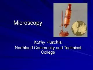
Microscopy. Core Concept # 1. Microscopy the study of objects or organisms (e.g., bacteria, protists, cells, etc.) too small to be seen by the naked eye using a tool called a microscope. Core Concept # 2. Microscope An instrument that gives an enlarged image of the object under study

Microscopy
E N D
Presentation Transcript
Core Concept # 1 • Microscopy • the study of objects or organisms (e.g., bacteria, protists, cells, etc.) too small to be seen by the naked eye using a tool called a microscope
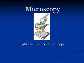
Core Concept # 2 • Microscope • An instrument that gives an enlarged image of the object under study • Two general types • Compound microscope • Electron microscope
Core Concept # 3 • Compound microscope • One of the basic laboratory tools of a microbiologist • Uses natural light to illuminate its specimen • Some have their own light bulbs for enhanced lighting
Core Concept # 4 • Compound: two lens systems • Primary lens system: objectives Secondary lens system: ocular/eyepiece • Based on the path of light (i.e., which lens system is hit by light first?) • The image is magnified twice
Rules in using the microscope • Proper handling • Always use BOTH hands: one on arm, the other under the base • Carry the microscope upright • Keep the microscope close to your body • Place the microscope on a dry, level surface, far enough from the edge of the table
Rules in using the microscope • Focusing • Under LPO • Optional: start with SO • Start with stage closest to the objectives • Use the coarse adjustment knob for initial focusing; use fine adjustment knob to sharpen the image • Under HPO • Once image is focused under LPO, DO NOT move the coarse adjustment knob anymore • Switch to HPO while looking from the side of the microscope (to make sure the HPO won’t hit the stage or slide) • NEVER use the coarse adjustment knob while using HPO
Rules in using the microscope • Preparing the specimens • Wet mount • Place specimen (thin enough for light to pass through) on a clean slide • Add 1-2 drops of water • Place a clean cover slip on the slide, starting with one edge then carefully lowering the rest of the cover slip • Staining • Place several drops of stain on one edge of the cover slip; the process of diffusion will allow the stain to go under the cover slip and stain the specimen (tissue paper may be placed on the opposite edge of the cover slip to help the stain diffuse under the cover slip) • Stain may be added directly to the specimen if it is not necessary to view the unstained specimen
Rules in using the microscope • Preparing the microscope for storage • SO in place • Mirror is parallel to the stage, with the plane side facing up • Stage is clean and dry and at the lowest position • Iris diaphragm is closed • Swivel screw is tightened
Computations in microscopy • Theoretical magnification = ocular X objective • Estimating the diameter of the field of view (LPO diam)(LPO mag) = (HPO diam)(HPO mag) • Estimating cell size = diameter / # of cells spanning the diameter
Lab Act # 3: Plant and Animal Cells • Onion (Alliumcepa) • Unstained, LPO • Stained w/ iodine sol’n • LPO • HPO
Lab Act # 3: Plant and Animal Cells • Hydrilla • Unstained • LPO • HPO
Lab Act # 3: Plant and Animal Cells • Cheek cells • Stained w/ methylene blue • LPO • HPO
(1/4 sheet) Bio1 3rd Qtr – Quiz # 2CN – Name DateSection • Given • LPO diameter = 2mm • Assume: microscope used is the same as that of the school’s (mirrored) • Find • Estimated average cell length • Show all important equations and the complete solution • Box your final answer Fig. 1. Onion cells stained with iodine solution under 400X