Download

1 / 28
630 likes | 2.84k Vues
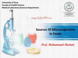
Detection Of Microorganisms In Food. Importance. The total microbial population in a food varies greatly and depends on: *The level of sanitation used at all phases. *The degree of abuse that leads to microbial growth.

E N D
Importance The total microbial population in a food varies greatly and depends on: *The level of sanitation used at all phases. *The degree of abuse that leads to microbial growth. *The processing and preservation methods used to kill and prevent growth of microorganisms. Contamination of a food by specific types or species of microorganisms depends on the presence of a source of these microorganisms, and their entrance into the food mostly due to poor sanitation during handling and processing.
Objectives *Microbiological examination of foods and food ingredients helps to * Assess their safety to consumers, *Their stability or shelf life under normal storage conditions and *The level of sanitation used during handling. In addition, the microbiological load and type can be important in determining whether a food or food ingredient meet acceptable standards, specifications and guidelines
Microbiological evaluation of raw materials also provides important information about the heat-processing parameters that would be necessary to meet the microbiological standards, guidelines, or specifications of a product. Microbiological evaluation of a food, food ingredient, and environment helps determine possible sources of a specific microbial type in a food and in the case of heated food, the source and nature of postheat treatment contamination.
Methods Used These methods are whether qualitative or quantitative. Quantitative methods are designed to enumerate or estimate directly or indirectly the microbial load in a test material. None of the quantitative methods used now enumerate or estimate total microbes, total bacteria, or total viable population, rather each method enumerates or estimates a specific group among the total microbial population normally present in a food and that grows or multiplies preferentially under the conditions or methods of testing. These include composition of an enumeration medium, temperature, time of incubation, oxygen availability, pH, and treatments of a sample before enumeration and estimation.
Examples of quantitative methods: Aerobic Plate Count(APCs),or standard Plate Count(SPCs) for dairy products. Anaerobic Counts, Psychrotrophic Counts, Thermoduric Counts, Coliform Counts, S.aureus Counts, Yeast and Mold Counts. Qualitative Methods: They determine whether a representative amount (a sample) of a food or a certain number of samples in a batch of a food contain a specific microbial species among the total microbial population.
Qualitative methods are used to detect the possible presence of certain foodbornepathogens, especially those capable of causing high fatality rates among consumers. Salmonella, Clostridium botulinum, Escherichia coli 0157:H7, and probably Listeria monocytogenes in ready-to-eat food, are some that fall into this group.
Standard and Recommended Methods *Sample Procedure The sample should be collected by using proper sanitary measures to prevent any contamination. Following collection and until tested, the samples should not be handled to avoid growth or death of microorganisms. If the product is frozen, samples should be kept frozen until analyzed. Otherwise, they can be stored at 0-4C. Each sample should be labeled to identify date, time, nature of sample, and types of analysis to be conducted, and the persons who collected the sample.
The samples should be transported to the laboratory under conditions that avoid microbial contamination, growth, or death. The laboratory that will test the samples should examine the conditions, such as temperature, appearance, and the sampling information, and note the time of receiving the samples. Once received in good condition, a sample should be tested as soon as possible. The unused portion of the sample should be stored under proper conditions until the results are available. The results should be recorded immediately and properly in permanent form. Standard or recommended methods should be used to prepare the sample and the procedures of testing for a specific microorganism or a Microbial group.
Quantitative Methods For Microbial Enumeration In Foods Colony-Forming Units (CFUs) in Nonselective Agar Media: Aliquotes from selected dilutions of a serially diluted sample are either pour plated or surface plated by using nonselective media such as plate count agar(PCA), tryptic soy agar, or nutrient agar. The temperature and time of plate incubation differ with the microbial groups. For SPCs, they are 32C for 48 h; APCs.35C for 48 h; for psychrotrophic counts, 7C for 10 days or 10C for 7 days.
The same procedures with specific modifications can be used to determine thermophilic, thermoduric, and anaerobic groups present in a food sample. The specific groups to be tested depend on their relative importance in a food. For a vacuum-packaged refrigerated food, the most important groups will be psychrotrophic, anaerobic, and facultative anaerobic groups.
CFUs in Nonselective Differential Media A nonselective medium is supplemented with an agent capable of differentiating the colonies produced by specific groups of microorganisms that differ in metabolic or physiological characteristics from one another in the population. pH indicators are often used in the medium. Colonies of cells capable of metabolizing lactose to lactic acid are differentiated from those that do not ferment lactose by growing them in agar medium supplemented with lactose as a carbon source and a pH indicator such as bromocresol purple. The lactose fermenting colonies will be yellow, others will be white.
CFUs in Selective Agar Media A medium can be supplemented with one or more selective or inhibitory agents and used by pour or surface plating of serially diluted samples. In the presence of such an agent, only the microorganisms resistant to it can grow. Incubation conditions to stimulate colony formation differ with the organisms being studied. Enumeration of aciduric bacteria in a medium at pH 5.0, yeasts and molds at pH 3.5, C. perfringensin the presence of cycloserine are examples of selective enumeration of specific groups in food. Halophilic bacteria can be enumerated by specific selective procedures.
CFUs in Selective-Differential Agar Media A medium is supplemented with one or more selective agents to allow selective growth of specific resistant microbial groups while inhibiting growth of other sensitive microbes. In addition to selective agents, a medium is also supplemented with agents that enable each type among the selective microbial groups to produce colonies that differ in characteristics from one another. Violet red bile agar for coliforms, KF-azide agar for Enterococcus spp. Are selective as well as differential agar media. Selective agent allows selective growth and colony formation, while differential agents help differentiate these species from one another by their specific colonies.
Indirect Estimation *Most Probable Number(MPN) in Selective Broths: Aliquots from a serially diluted sample are inoculated in a broth(in tubes) having one or more selective agents that facilitate growth of selected microbial groups present in a food. Three or five broth tubes in each dilution and a minimum of three consecutive dilutions are used. After incubating at recommended temperature and time, the broth tubes in each dilution are scored for the presence and absence of growth.
From the number of tubes showing growth in each of the three successive dilutions, the number of viable cells of the specific microbial group can be estimated from the available statistically calculated tables. This method gives wide variation. MPN methods are used to estimate coliforms and fecal coliforms in foods and water by using brilliant green lactose bile broth and EC broth.
Dye Reduction Test Some dyes such as methylene blue and resazurin are colored in oxidized states but colorless under reduced conditions. This change can occur because of microbial metabolism and growth. It is assumed that the rate of reduction during incubation of a specific concentration of methylene blue added to a food is directly proportional to the initial microbial load in the food. This method is generally used to determine the microbiological quality of raw milk.
Qualitative Methods Isolation of Pathogens The main objective of this method is to determine whether a sample contains viable cells or spores of a specific pathogen. Foods are tested for several pathogens, such as Salmonella, E.coli 0157:H7, L. monocytogenes, Vibrio cholerae, and Shigella spp. By the specific isolation procedure. For other pathogens, such as enteropathogenicE. coli, Y.enterocolitica, and campylobacter jejuni, isolation procedures are not generally used, instead, enumeration procedures are used.
An isolation procedure contains several steps: *nonselective preenrichment *Selective enrichment *Testing on an agar medium containing selective and differential agents. A food normally contains a low population of a pathogen as compared with associative microorganisms, and the pathogens could be in the injured state. The food sample(e.g., 25 g) is first preenriched in a nonselective broth and incubated for the injured cells to repair and then multiply in order to reach moderately high numbers.
An aliquot is then transferred from the preenrichment broth to a selective enrichment broth and incubated. It is expected that during incubation, the specific pathogen and closely related microorganisms will selectively grow to a high number, whereas many of the associated microorganisms will not grow. A small amount(0.01 ml) of the enrichment broth is streaked on the surface of a prepoured selective-differential agar medium plate, which is then incubated for specified time for the colonies to develop. The presence of a specific pathogen can be tentatively established from the colony characteristics.
This is generally considered a presumptive test. For confirmation, the cells from characteristic colonies are purified and examined for biochemical reaction profiles and serological reaction against specific antibody. Isolation of a pathogen using the conventional methods can take 10-12 days, depending on a particular species.
Test For Bacterial Toxins In Foods *S. aureus Enterotoxin Toxin is extracted from 100 g food, then it is concentrated in 0.2 ml M saline. Toxin is detected by reaction with specific antibody on a microslide. ELISA can also be used. *Botulin Toxin The toxin is extracted from food, then it is activated by trypsin treatment, 0.5-ml is injected into mice intraperitoneally, control(a portion is heated at 100C) is also injected into another mice. The mice are observed for botulism symptoms and death.
Rapid Methods for Detection of Microorganisms in Food The conventional methods used for the quantitative or qualitative detection of microorganisms and toxins in foods take a relatively long time. different rapid methods have been developed To detect microbial loads, foodborne pathogens, and their toxins. In addition to being rapid, they are specific, sensitive, accurate, and less labor intensive.
Immunofluorescence Specific fluorescence-conjugated antibodies which are directed against somatic or flagellar antigens of a pathogen are mixed with an enriched medium suspected to contain the specific pathogen, such as Salmonella, on a glass slide. Following incubation and removal of reagents, the slide is examined under a fluorescence microscope for cell showing fluorescence on the cell wall or flagella or both.
Enzyme Linked Immunosorbent Assay(ELISA) The specific antibody is allowed to bind on a solid surface(microtitration plastic plate). The sample suspected of containing the antigen(pathogens or their toxins) is prepared and added to the well and incubated for the antibody-antigen reaction. After removing the unbound antigen(washing step), another antibody labeled with a specific enzyme(such as alkaline phophatase) is added and incubated for antigen binding, a sandwich is formed(Ab-Ag-Ab*enzyme).
The unbound enzyme-linked antibody is then removed. The sandwich complex is detected by adding a chromogenic substrate specific for the enzyme(such as p-nitrophenyl phosphate), incubating for a specified time , and adding an enzyme inactivator to stop the reaction. The intensity of the color can then be measured to identify the presence of a specific pathogen or toxin.
Polymerase Chain Reaction(PCR) The PCR technique helps amplify a segment of DNA. It is thus possible to obtain large numbers of copies of a specific DNA segment from a small sample(50 ng), which in turn facilitates its detection by gel electrophoresis. PCR is a sensitive method and may not need preenrichment or enrichment steps. To eliminate confusion between dead and viable cells of a pathogen as the source of DNA, a short preenrichment step may be necessary to increase the viable cell number of the target pathogen.
Bioluminescence The bioluminescence method measures the ATP content in a sample as an indirect measurement of microbial load. As only viable cells retain ATP, the amount of ATP is regarded as directly related to the microbial load in the sample. Using the luciferin-luciferase(from firefly) system in the presence of Mg, the ATP concentration in the lysed cells in a sample is measured. The method is very rapid.