High content screening workflow
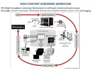
High content screening workflow. HTS (High throughput screening): Biochemical or cell based; mostly end-point assays HCS (High content screening): Cell based; fluorescence imaging; kinetic output / live cell imaging. High Content Screening (HCS) applications :

High content screening workflow
E N D
Presentation Transcript
High content screening workflow HTS (High throughput screening): Biochemical or cell based; mostly end-point assays HCS (High content screening): Cell based; fluorescence imaging; kinetic output / live cell imaging
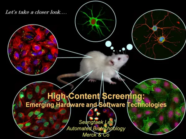
High Content Screening (HCS) applications: • Target identification - Genome-wide functional siRNA screening. • Primary screening - Screening of libraries of compounds with meaningful functional cellular readout. • Secondary screening / follow up testing - Dose responses for active compounds, complementary assay to identify false positives. Time courses of the assay in live cells. • Safety assessment: In vitro tests using organ specific cells (e. g. hepatocytes, neurons) in order to get early information on the side effects of drugs. • Systems biology: Genome-wide functional siRNA screening. Recent advances in yeast biology, i. e. the availability of genome-wide collections of gene-knockout mutants, has greatly facilitated yeast-based gene function discovery. • Stem cell biology: The search for stem cell based therapies and scalable human primary cell sources for drug discovery have created an enormous need for stem cell reprogramming and differentiation screening methods.
High content screening platforms and categories • Thermo Fisher/Cellomics • Cytotoxicity and apoptosis • Genotoxicity, DNA damage and repair • Inflammation and Cell Stress • Cell Signaling and Transcription Factors • Cell Cycle and Proliferation • Cell Morphology and Phenotype Changes • Perkin Elmer • GE Healthcare • Molecular Devices • Genetix • BD Biosciences
Specifications of high content screening analysis • Assay features • Assay category • Assay name • Target • Biological process • Potentiator • Pathway/Target • Species • Cell-line • Phenotype • Detection • Primary antibody • Secondary antibody • Dye • Dye target • Image analysis • Image analysis module • Detection target • Detection quantification • Endpoint • Controls • Data normalization
Example specifications • Assay features • Assay category: Cytotoxicity and apoptosis • Assay name: Apoptosis 1 • Target • Biological process: Apoptosis • Potentiator: Doxorubicin • Pathway/Target: Mitochondrial pathway of apoptosis • Species: Human • Cell-line: Hek-293 • Phenotype: Nuclear DNA condensation • Detection • Primary antibody: Caspase-3 • Secondary antibody: Anti-caspase-3 conjugated with Alexa 594 • Dye: Hoechst • Dye target: DNA • Image analysis • Image analysis module: Cell health profiling bioapplication • Detection target: Nuclear DNA • Detection quantification: Fluorescence intensity • Endpoint: Concentration response; activity based; generic • Controls: DMSO negative control • Data normalization: Z’-factor
High content analysis • Application • Channel 1 • Cell counts and density • Output cell features: e.g. Cell number • Output well features: e.g. ValidObjectCount • Morphology and location • Output cell features: e.g. ObjectArea and Status • Output well features: e.g. Mean, SD • Intensity • Output cell features: e.g. Total intensity and status • Output well features: e.g. Mean, SD • Channel 2 • Intensity • Output cell features: e.g. Total intensity and status • Output cell features: e.g. Mean, SD • Morphology • Output cell features • Output cell features
Existing Cell line ontologies Cell line MCCL: http://bioportal.bioontology.org/visualize/39927 Cell line CLKB: http://bioportal.bioontology.org/visualize/40261 ATCC has 3487 and CLDB has 5367 cell lines Existing fluorophore ontologies Molecular Imaging and Contrast Agent Database (MICAD): http://www.ncbi.nlm.nih.gov/bookshelf/br.fcgi?book=micad Biological imaging methods: http://www.ebi.ac.uk/ontology-lookup/browse.do?ontName=FBbi
Cell line MCCL ontology • Anatomical entity • animal kingdom • cell • cell in vivo • experimentally modified cell • cell line cell • permanent cell line cell • 1321N1 • 16610D9 • 16HBE14o- • 1HAEo • 2008/C13*5.25 • 293 • 2fTGH • 308 • 30A5 • 32D • 32Dcl3 • 3T3-Swiss albino
Molecular Imaging and Contrast Agent Database (MICAD) OpticalA15 Alexa Fluor 488 Alexa Fluor 647 Alexa Fluor 680 Alexa Fluor 750 AOI987 Au BODIPY-FL BODIPY TMR-X Cy5 Cy5.5 Cy7 Cypate DiD DiR Fluorescein Fluoresceinisothiocyanate (FITC) Green fluorescence protein Green fluorescence protein (GFP), luciferin HiLyte Fluor 647 Indocyanine green IR-783 IR-786 IRDye700DX IRDye78 IRDye 800CW Luciferin Malachite green NIAD-4 NIR2 Oregon Green 488 Pyropheophorbideα (Pyro) Quantum dot (QD) Rhodamine green (RhodG) RhodamineX SIDAG Single-walled carbon nanotubes (SWNTs) Tetramethyl-6-carboxyrhodamine VivoTag-S680 VM315