Download
1 / 38
390 likes | 681 Vues
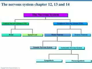
. The Central Nervous System. CNSBrainSpinal cord ~. Peripheral Nervous System. Somatic Nervous System - SNSSensory

E N D
1. Anatomy of the Central Nervous System:Part 1 Lecture 10
3. Peripheral Nervous System Somatic Nervous System - SNS
Sensory & Motor
Autonomic Nervous System - ANS
Sympathetic Division
Parasympathetic Division
Cranial Nerves - 12 Pair
Spinal Nerves - 31 Pair ~
4. Terminology Afferent - toward
Sensory
Efferent - away from
Motor ~
5. Dorsal - toward the back
Ventral - toward the stomach
Anterior & Posterior
Rostral & Caudal ~ Terminology
7. Brainstem & spinal cord
10. Terminology Medial - toward the midline
Lateral - away from the midline
Ipsilateral - same side
Contralateral - opposite side
Decussate - crossover ~
11. Coronal - frontal
cross-section - salami - front view
Sagittal - side view
hot-dog bun
Horizontal - top view ~ Planes
15. Development of the Nervous System
16. Embryological Origin Embryo has 3 layers
endoderm ---> internal organs
mesoderm ---> muscles & skeleton
ectoderm ---> nervous system & skin
Neural plate (~3 weeks)
sheet of cells
neural groove
folds rostral to caudal ---> tube ~
17. Development of N.S. Neural tube
CNS develops from walls
as tube develops dorsal portion pinches off & forms...
Neural crest
develops into all neurons of PNS
Mesoderm
somites ---> vertebrae & muscles ~
18. Neural Tube Defects Neural tube fails to close
Rostral failure
anencephaly
skull & brain partially or totally absent
perinatal mortality
Encephalocele
parts of brain protrude outside skull
mental disability depends on extent ~
19. Neural Tube Defects: Spina Bifida Cleft spine
Caudal neural tube fails to close
Incidence
Spina bifida occulta
40% of Americans
Spina Bifida Manifesta
1 in a 1000 births
myelomeningocele: 96%
meningocele: 4% ~
20. Neural Tube Defects: Spina Bifida Myelomeningocele
protruding cyst as with meningocele
spinal cord / nerves in protruding sac
Effects
paralysis, incontinence, learning disabilities
70-90% also hydrocephalus
can cause severe brain damage
Repaired surgically ~
21. Neural Tube Defects: Spina Bifida Meningocele
fluid-filled meningeal cyst
protrudes through unfused vertebral arches
SC/nerves not in sac
repaired surgically
less severe disability than myelomeningocele ~
22. Neural Tube Defects: Prevention Possible genetic component
reoccurrence in families
ethnic/racial relationship
Folic Acid (Folate) Deficiency
50-70% of cases preventable
CDC guidelines
not planning pregnancy: 400 mg/day
planned pregnancy: 4000 mg/day
under direction of health care provider ~
23. Stages of Cellular Activity 6 distinct stages
1. Neurogenesis
mitosis
nonneural cells
neurons do not divide
develop into neurons or glia
glia produced throughout life ~
24. Stages of Cellular Activity 2. Cell migration
to specific �brain� locations
along radial glia
later: along other neurons
3. Differentiation
into specific neuron types
by induction
influenced by surrounding cells
or cell-autonomous ~
25. Stages of Cellular Activity 4. Synaptogenesis
formation of synapses
growth of axons & dendrites
growth cones
5. Neuronal cell death
apoptosis: programmed cell death
20-80% of neurons in a region
genetically programmed
lack of neurotrophic factors ~
26. Stages of Cellular Activity 6. synaptic rearrangement
elimination of synapse
formation of new synapses
dependent on neural activity ~
27. Differentiation Specialization of structures
3 primary vesicles
rostral end of tube
develops into brain
Prosencephalon ---> forebrain
Mesencephalon ---> midbrain
Rhombencephalon ---> hindbrain ~
28. Prosencephalon Secondary vesicles form & separate
optic ---> retinas
retina & optic nerve part of brain
not PNS
telencephalic ---> telencephalon
remainder ---> diencephalon ~
29. Other Primary Vesicles Mesencephalic ---> mesencephalon
dorsal - tectum
ventral - tegmentum
tube - cerebral aqueduct
Rhombencephalic
rostral - metencephalon
caudal - myelencephalon
tube - 4th ventricle ~
30. Telencephalon Cortical areas
Fiber systems
Landmarks
Fissures /Sulci
Lobes
Gyri ~
35. The Meninges Tough connective tissue
Dura mater - tough outer layer
Arachnoid membrane - soft middle layer
Pia mater - soft inner layer, next to brain ~
36. Subarachnoid space Gap between arachnoid membrane and pia mater
Filled with CSF
Brain floats in CSF ~
37. Dura mater
Arachnoid membrane
Subarachnoid space CSF
Pia mater
The Meninges
38. Ventricular System Another circulatory system for the brain
4 interconnected ventricles
chambers
Cerebral Spinal Fluid (CSF)
manufactured in choroid plexus
Hydrocephalus - blockage of CSF flow ~
39. Ventricular System