Download
1 / 16
160 likes | 291 Vues
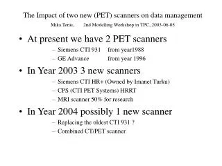
The Impact of two new (PET) scanners on data management Mika Teräs, 2nd Modelling Workshop in TPC, 2003-06-05. At present we have 2 PET scanners Siemens CTI 931 from year1988 GE Advance from year 1996 In Year 2003 3 new scanners Siemens CTI HR+ (Owned by Imanet Turku)

E N D
The Impact of two new (PET) scanners on data managementMika Teräs,2nd Modelling Workshop in TPC, 2003-06-05 • At present we have 2 PET scanners • Siemens CTI 931 from year1988 • GE Advance from year 1996 • In Year 2003 3 new scanners • Siemens CTI HR+ (Owned by Imanet Turku) • CPS (CTI PET Systems) HRRT • MRI scanner 50% for research • In Year 2004 possibly 1 new scanner • Replacing the oldest CTI 931 ? • Combined CT/PET scanner
PET Det. Mat. Rings = n (--> 2n-1 slices) Cryst/Ring Crystal Dim. mm Cryst/Block Slice thick ness mm Axial FOV cm GE Advance BGO 18 672 4.0x8.1x30 6 x 6 4.25 15.2 ECAT 931 BGO 8 512 5.6x13x30 4 x 8 6.75 10.8 HRRT LSO/LYSO 104 104 576 576 1.9x1.9x10 1.9x1.9x10 8 x 8 8 x 8 1.2 24.0 ECAT HR + BGO 32 576 4.0x4.4x30 8 x 8 2.5 15.5 PET Tomographs
PET Trans axial res. mm Sensi-tivity cps/Bq/ml NEC kcps Sinogram dimension Raw data size Mb Axial FOV (cm) GE Advance 5 160 281x256 50 15.2 ECAT 931 7 5 50 192x336 1.6 10.8 HRRT 3 35 220 256x288 x2 Aprox. 500 24.0 ECAT HR + 5 31 110 288x144 20 15.5 PET Tomographs 2 31
Issues to be handled 1 • new data formats • HRRT: Interfile, Flat Format • HR+: ECAT 7.n • MRI: not yet known (DICOM) • raw data archiving • local DAT drives as now or some centralised system • reconstruction requirements • standard reconstruction • iterative reconstruction • research for new methods
HRRT data organization and size HRRT “Ring 1-Ring 2” coincidence map All data are stored in flat format + Interfile header. Standard acquisition mode is (span 9, rd 67). It corresponds to 2209 sinograms in 15 segments. Max. co-polar angle is ≈ 19° Scan size (256x288x2209x2) is 325 MB. Norm/Atten size are both 650 MB. Image (256x256x207x4) size is 54 MB/frame. At high resolution (span 3, rd 67), we have 6367 sinograms in 43 segments: data size is increased by 2.9 compared to std mode !!! z span segment -7 q Every dot in the map is a sinogram (256 x 288) => (104)2 sinograms
HRRT Interfile header: example for a scan file number of bytes per pixel := 2 image relative start time := 720 frame := 1 Total Prompts := 765518378 Total Randoms := 295242655 Total Net Trues := 470275723 block singles 0 := 13456 block singles 1 := 14296 block singles 2 := 15965 block singles 3 := 14428 block singles 4 := 16499 block singles 5 := 16442 block singles 6 := 17529 block singles 7 := 13435 block singles 8 := 10770 .... block singles 934 := 18109 block singles 935 := 17051 average singles per block := 22714 Dead time correction factor := 1.191135 !originating system := HRRT !name of data file := g:\sinogram\lenox-mark-27335-2002.l32 !study date (dd:mm:yryr) := 14:03:2002 !study time (hh:mm:ss GMT) := 15:57:12 !PET data type := emission data format := sinogram number format := short number of byte per pixel := 4 axial compression := 9 maximum ring difference := 67 energy window lower level[1] := 350 energy window upper level[1] := 650 image duration := 600 Patient name := lenoxCTI, mark Patient DOB := 12:00:00 AM Patient ID := 11111 Patient sex := Male Dose type := FDG Dosage Strength := 2.1 Mega-Bq Normalization file name and path := G:\norm\default.n Blank file name and path := g:\blank\default.s matrix size [1] := 256 matrix size [2] := 288 matrix size [3] := 2209 Singles
Issues to be handled 2 • image data flow • new analysis workstations for clinical data • new MRI data arrangement • new security aspects • Imanet Turku Studies • compatibility with modelling programs • new image format (ECAT7 ?) • HR+ compatible with the Imanet London scanner • acquiring of all related data • motion correction with Polaris on List Mode data • blood and activity measurements with new systems • continuous blood sampling • metabolite analysis
ECAT EXACT HR+: Premium Performance • Highest overall performance scanner available for PET • Finest sampling (2.4 mm) • Highest resolution • High image quality
ECAT to e.soft Biograph console Sun Acquisition Console e.soft Database Browser
e.soft PET Display • DICOM connectivity • Configure display • Zoom • Autonormalize color scale • Add slices • Dual volume display • View MIPs • Draw ROIs (SUVs)
HRRTHardware & software improvements ... • New front end electronics and new light tight interface: improves reliability (less power inside head) and easier access for service. • The integration time was reduced to 120ns with subsequent improvements in countrate performance • Faster acquisition hardware was introduced (2.0 Ghz Intel) which achieves higher histogramming rates (6.5 Mevents/s) with a larger memory footprint (2.0 GB) • A 100 MB/sec 1 TB RAID allows storage of listmode data at high rate for long periods of time.
Acquired in list mode with a rotating rod: long, uniform, weak. Measurement statistics: as long as possible (week-end) Fit data for rod radius and center of rotation, calculate dwell-time for transforming ring into plane source. Invert data. Not component-based: 1 file per acq mode: (9,67) is 650 MB. Is used to detect missing data for reconstruction HRRT Normalization 2 D segment of 3D normalization
HRRT OSEM3D on the Windows/NT Heinzel Cluster • A parallel version of OSEM3D code has been implemented on a farm of 7 SGI quad-processors (28 Pentium III Xeon@700 Mhz). It uses both shared and distributed memory and Myrinet networking hardware with the GM Message Passing System. [S. Vollmar et al, IEEE-MIC 2000 & MIC 2001 and 3D 01 conferences].
HRRT PET-MR fusion at high PET resolution ... If we want to navigate into PET using inverse gray, the alternative is to transform the MR CLUT into an edge detector and fuse these contours with PET.
Intsalled at the end of 2001 DISCOVERY Intsalled April 2002 REVEAL HD
Whole-body PET CT PET PET Reconstruction RHDDLS+ FORE + 2D-OSEM2D-OSEM