Download

1 / 34
510 likes | 2.96k Vues
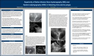
Saline Infusion Sonohysterography. Robert D. Auerbach, M.D. FACOG Senior Vice President & Chief Medical Officer CooperSurgical, Inc. Associate Clinical Professor Yale University School of Medicine. Introduction. Saline Infusion Sonohysterography (SIS) Transcervical introduction of fluid

E N D
Saline Infusion Sonohysterography Robert D. Auerbach, M.D. FACOG Senior Vice President & Chief Medical Officer CooperSurgical, Inc. Associate Clinical Professor Yale University School of Medicine
Introduction • Saline Infusion Sonohysterography (SIS) • Transcervical introduction of fluid • Provides enhanced endometrial visualization during TV U/S examination • Benefits • Easy to perform • Minimal cost • Well tolerated by patients • Few complications
Indications & Contraindications • Indications • Abnormal Uterine Bleeding • Infertility and habitual abortion • Congenital abnormalities of the uterus • Pre or postoperative evaluation of uterine myomas, polyps, cysts • Suspected uterine cavity synechiae • Inadequate TV U/S
Indications & Contraindications • Contraindications • Pregnancy • Pelvic infection AIUM standard for the performance of Saline Infusion Sonohysterography www.aium.org/consumer/standards/saline.pdf
Indications - AUB • Abnormal Uterine Bleeding (AUB) • Accounts for many patients visits • Diagnostic procedures • TV U/S • Endometrial Biopsy • Hysteroscopy • D&C – no longer considered an initial diagnostic test
Indications - AUB • SIS is useful for finding focal lesions in AUB • 106 patients with menometrorrhagia * Significant difference and comparable to hysteroscopy Dx rates Kamel, HS, Darwish, AM, Mohamed, SA. Comparison of transvaginal ultrasonography and vaginal sonohysterography in the detection of endometrial polyps. Acta Obstet Gynecol Scand 2000; 79:60.
Indications - AUB De Kroon, CD, Jansen, FW, Louwe, LA, et al. Technology assessment of saline contrast hysterosonography. Am J Obstet Gynecol 2003; 188:945. • Prospective series of 214 consecutive pre- and postmenopausal women with AUB • Suspected uterine abnormality on TV U/S • SIS followed by hysteroscopy preferable to hysteroscopy initially • Hysteroscopy could be avoided 84% of patients
Indications - AUB • SIS can help distinguish: • Anatomic AUB from anovulatory bleeding • Postmenopausal bleeding from atrophy • May help select appropriate candidates for endometrial sampling • Differentiate focal lesions that need biopsy under direct vision versus a global process that can be sampled blindly
Indication - Infertility • Assessing the endometrium is part of the routine infertility & recurrent pregnancy loss work-up • SIS can Dx anatomic causes of infertility • Submucous myomas • Endometrial polyps • Uterine anomalies • Intrauterine adhesions
Indication - Infertility • SIS is comparable or better than HSG • Salle, B, Gaucherand, P, de Saint, Hilaire P, Rudigoz, RC. Transvaginal sonohysterographic evaluation of intrauterine adhesions. J Clin Ultrasound 1999; 27:131. • Darwish, AM, Youssef, AA. Screening sonohysterography in infertility. Gynecol Obstet Invest 1999; 48:43. • Alborzi, S, Dehbashi, S, Parsanezhad, ME. Differential diagnosis of septate and bicornuate uterus by sonohysterography eliminates the need for laparoscopy. Fertil Steril 2002; 78:176. • Alatas, C, Aksoy, E, Akarsu, C, et al. Evaluation of intrauterine abnormalities in infertile patients by sonohysterography. Hum Reprod 1997; 12:487. • HSG better at outlining fallopian tubes
Indication - Tamoxifen • Tamoxifen is used extensively as adjunctive chemotherapy in women with estrogen receptor-positive breast cancer • May thicken the uterine wall in some women when viewed by TV U/S • May be polyps or abnormal endometrial tissue
Indication - Tamoxifen • SIS has shown these changes are often microcysts of the basalis (focal adenomyosis reactivation) • Hann, LE, Gretz, EM, Bach, AM, Francis, SM. Sonohysterography for evaluation of the endometrium in women treated with tamoxifen. AJR Am J Roentgenol 2001; 177:337. • Elhelw, B, Ghorab, MN, Farrag, SH. Saline sonohysterography for monitoring asymptomatic postmenopausal breast cancer patients taking tamoxifen. Int J Gynaecol Obstet 1999; 67:81. • Routine sonographic screening of the endometrium of asymptomatic women taking Tamoxefen has not been recommended by ACOG
SIS Technique • Usually scheduled between day 5 – 10 of the menstrual cycle • No routine anesthesia or analgesia needed • Bimanual exam • Place speculum • Aseptic prep of the cervix • SIS • Flush catheter • Insert catheter • May be accomplished without the need for a tenaculum
SIS Technique • Fill balloon and snug catheter against the internal os • Remove speculum • Attach 10ml syringe of sterile saline • Insert TV U/S probe
SIS Technique • Fluid is instilled while the probe is shifted to each cornual region • Long axis plane • Turn probe 90 degrees and instill more fluid • Coronal plane • Fan from the endocerival canal to the fundus
Findings - Polyp TV US for abnormal bleeding revealed an ill-defined mass SIS demonstrates fundal polyp
Polyp - Removal Polyp removed under U/S guidance leaving a normal endometrial cavity (left)
Multiple Polyps Multiple polyps in a 52-year-old woman with abnormal bleeding. Saline infusion sonohysterography shows multiple polypoid masses (arrows) outlined by the anechoic saline.
Cancerous Polyps Papillary serous carcinoma in a 71-year-old woman with postmenopausal bleeding. Findings from blind endometrial biopsy performed before the SIS were negative.
Findings – Submucosal Myoma TV US shows myoma with non-distinct borders SIS shows myoma protruding into endometrial cavity
Findings - Tamoxifen TV US showing irregular endometrium and cysts SIS showing thin endometrium and sub-basalis cysts
Endometrial Hyperplasia Saline infusion sonohysterography shows diffuse endometrial thickening and multiple cystic areas (arrowheads). Pathologic examination showed simple endometrial hyperplasia.
Endometrial Hyperplasia Endometrial hyperplasia in a 54-year-old woman with menorrhagia showing areas of focal thickening of the endometrium. Pathologic examination revealed complex endometrial hyperplasia without atypia.
Findings – Endometrial Cancer TV US shows 7mm endometrium after menses – should be no > than 4mm SIS demonstrates inapprop. thick endometrium – biopsy reveals well differentiated adenocarcinoma
Metastatic Cancer Mass in a 71-year-old woman with a history of breast cancer treated with tamoxifen for approximately 2 years who had PMP bleeding. SIS shows an inhomogeneous focal mass (arrows). Pathologic examination revealed metastatic breast cancer to the uterus.
Findings - Adhesions Intrauterine adhesions in a 33-year-old woman undergoing a fertility workup with a history of 2 dilation and curettage procedures. Saline infusion sonohysterography shows a poorly distensible endometrial cavity with adhesions (arrows).
Technical Difficulties & Complications • Passing the catheter • Cervical stenosis • Uterine position • Uterine abnormality • Poor visualization • Unable to retain fluid (patulous cervix) • Infection • Same risk as HSG • Obtain cultures and Rx with AB based on risk factors
Technical Difficulties & Complications • Cancer dissemination – positive cytology equals at least Stage IIIA • Theoretic concerns • HSG study with spill survival rates unchanged • Devore, GR, Schwartz, PE, Morris, JM. Hysterography: a 5-year follow-up in patients with endometrial carcinoma. Obstet Gynecol 1982; 60:369. • False positive • Attributed to blood clots, intrauterine debris, mucus, shearing of the endometrium and thickened endometrial folds
Getting Fooled Inadvertent injection of a small amount of air. The air appears as a bright echogenic focus in the fundal region of the endometrial cavity
Getting Fooled Blood clot mimicking a mass. The patient had postmenopausal bleeding. Initial images (A and B) show an echogenic mass in the endometrial cavity (arrow). Color Doppler imaging (C) shows no color flow within the mass. During the examination, the mass was dislodged with the catheter, and the final image (D) shows a normal thin endometrium. C A D B
Technical Difficulties & Complications • Prospective study of 1153 women age 23-64 undergoing SIS • Failure to complete procedure: 7% • Pelvic pain: 3.8% • Vagal response: 3.5% • Nausea: 1% • Post procedure fever: 0.8% Dessole, S, Farina, M, Rubattu, G, et al. Side effects and complications of sonohysterosalpingography. Fertil Steril 2003; 80:620.
Summary • SIS provides enhanced endometrial visualization • Improves sonographic detection of polyps, hyperplasia, cancer, myomas and adhesions • Can help avoid invasive procedures • May optimize preoperative evaluation who require therapeutic intervention