Download
1 / 13
130 likes | 140 Vues
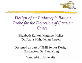
Design of an Endoscopic Raman Probe for the Detection of Ovarian Cancer. Elizabeth Kanter, Matthew Keller Dr. Anita Mahadevan-Jansen Designed as part of BME Senior Design (Instructor: Dr. Paul King) Vanderbilt University. The Problem: Ovarian Cancer.

E N D
Design of an Endoscopic Raman Probe for the Detection of Ovarian Cancer Elizabeth Kanter, Matthew Keller Dr. Anita Mahadevan-Jansen Designed as part of BME Senior Design (Instructor: Dr. Paul King) Vanderbilt University
The Problem: Ovarian Cancer • Fifth leading cause of cancer death among U.S. women • An estimated 25,400 U.S. women will be diagnosed with the disease, and an estimated 14,300 will die from it, in 2003 • Currently, 50 percent of the women diagnosed with ovarian cancer die from it within 5 years • 3.6 times more likely to develop ovarian cancer if have primary relative afflicted Source: National Cancer Institute and http://www.ovariancancer.org/content/1-5-1.html
The Problem: Ovarian Cancer • When detected before it has spread beyond the ovaries, there is a greater than 90 percent five-year survival rate • Late-stage diagnosis, which occurs in 75% of cases, causes an additional health care cost of approximately $40,000 over a patient’s lifetime • Difficult to diagnose because it is often asymptomatic, any symptoms present are easily confused with other diseases Source: National Cancer Institute and http://www.hcfinance.com/dec/dectside2.html
Motivation • There is currently no reliable, easily administered method for early detection of ovarian cancer • Optical spectroscopy has been proven to be effective as a real-time, non-intrusive, automated diagnostic tool for several physiological systems: • Colon, cervix, skin, esophagus • Raman spectroscopy has been shown to quickly give accurate biochemical information necessary to diagnose cancer Source: Mahadevan-Jansen (2002)
Raman Scattering & Spectra • Photons collide inelastically with scattering molecule • Molecule enters virtual excited vibrational state, then returns to lower state • Photon of lower frequency re-emitted • Raman Spectrum is plot of signal intensity vs. shift in wavenumber (1/wavelength) • Very weak signal, compared to fluorescence • Peaks narrow and highly specific to particular chemical bonds, which allows differentiation between normal & cancerous tissue
Overall System Scheme • 785 nm diode laser • Fused-silica low –OH fiber • Cooled CCD camera • Signal processing software
Ovarian Raman Spectra • Protein peaks are located at 1450 cm-1 and 1650 cm-1 • A DNA peak is located at 1330 cm-1. • Both protein and DNA peaks are increased in magnitude in cancerous tissue due to enlarged nuclei
Probe Constraints The in vivo Raman probe to be designed should • Fit inside the microlaparoscope, which has an inner diameter < 3 mm • Be able to visualize location of probe tip • Read only the Raman signal of tissue • Be in direct contact with tissue during each measurement • Not induce a harmful reaction in the body
Probe Design • ½ inch (12.7 mm) diameter optics • Bandpass filter: 785 +/- 5 nm • Longpass filter: OD of 6 for wavelength < 790 nm • CaF2 lenses • 400 μm core excitation fibers • 100 μm core collection fiber bundle with 11 fibers
Probe Testing (below) The first spectrum taken with the developed Raman probe (above) The probe being tested in the lab for the first time
One-time cost for clinics – approximately $2100 plus labor for probe itself Save money on future health care costs through reliable early detection Unique Minimally invasive – microlaparoscope < 3 mm in diameter, so leaves no scar and can be done with local anesthetic Benefits of Our System
Future Goals • Replacement of wrong lens and realignment • Extensive in vitro testing of probe • Statistical comparisons with other previously proven probes for analogous procedures • Eventually conduct clinical trials, although perhaps with a later generation model