Download
1 / 39
390 likes | 413 Vues
Explore the potential of Compton Back Scattering (CBS) sources for generating high-quality X-rays, enabling various applications from medical radiography to cultural heritage preservation. Learn about the efficiency and versatility of Compact Compton Sources (CCS) compared to synchrotron sources, paving the way for innovative research in diverse fields.

E N D
Alessandro Variola (LNF INFN) alessandro.variola@lnf.infn.it Compton sources for g and x ray applicationsWork supported by the EQUIPEX program, the Ile de
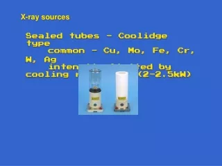
Compton backscattering sources • Compton Back Scattering (CBS) • Counter propagating electron and lasers beams. • Photon backscattering • Why CBS sources ? • CBS is by far the most efficient photon energy amplifier : wdiff=4g2wlaser, for example => g~100 => it is possible to have at one’s disposal hard X rays with a relatively low energy electron machine. • But for a light source: s ~ 6.6524 10-25 cm2 , it is low!!!!! • Need of a lot of photons and electrons, depending form the considered configuration for instantaneous or average brillance. • CBS attractiveness : • 1) Directivity (relativistic boost) = > f= 1/g around the electron direction • 2) Energy angle dependence => monochromatic by diaphragm • 3) Polarized if needed • 4) Backscattered spectrum cut off => Energy dependence on collision angle
1rst interest: the energy boost • (no polar. are observed) Energy distribution ~flat with w2,max=4g2wlaser with g~100 (Eelectron=50MeV) w2,max=45000eV if wlaser~ 1eV Compton scattering is the most powerful mechanism to boost photon energies The cut off is dependent on the incidence angle !! (factor two up to p/2) • Sprangle et al. JAP72(1992)5032
collimator wf(keV) q(mrad) • 2nd interest: the angular energy correlation • Compton scattering • Photon_laser+e photon+e’2 body process wf = f(q) • Sprangle et al. JAP72(1992)5032 • ~monoenergétic beam by selecting Backscattered photons at wf,max • Eelectron=50MeV
RMS bandwidth, due to collection angle, laser phase space distribution and electron beam phase space distribution • laser • electron beam • Diaphragm • L.Serafini
Applications of Compton scattering: quasi monochromatic X/g ray beam • Compact sources for high energy gammas g~100 MeV g ~1MeV • Elec.~20-100MeV • Elec. 1GeV • Elec.~100-750MeV wf,max MeV • X ray ~10-100keV • High energy applications • Compton polarimeterLEP energy measurement • Laser wire • gg collider • Polarised positron source • Low energy applications • Medical: radiography &radiotherapy • Museology • Material science • crystallography • Nuclear fluorescence applications • Nuclear survey • Nuclear waste management • Nuclear science
►In many scientific domains synchrotron sources are currently the only machines in term of brightness to perform and carry out the most ambitious analyses and searches requiring ~ 10-100 KeV X-rays. ► Synchrotron sources : - very powerful, but, - not very “pratical” for some applications, - limited access time. ► With Compact sources : Methods currently used at synchrotrons (diffraction, absorption, diffusion, imaging, spectroscopy…) could be largely developed in a laboratory size environment (hospitals, labs, museums). ‘compact’ source for nuclear physics photons (MeV range) -Nuclear safety -Nuclear waste management -Radioisotopes detection
Compton and other sources, X rays • ►X-ray tubes • - The most efficient are rotating anodes • - Rigaku ~ 1010 ph/sec , polychromatic • ►Plasma sources • Ultra-short pulses ~ fs , • but very low fluxes. • CCS • These sources does not allow to carry out • many of the techniques used at synchrotrons • ► Compact Compton Sources (CCS) • Compactness ( surface ~ 100 m2 ) • (Integration in hospitals, labs, museums) • Relative high intensity (1012 – 1014 ph/sec) • Tunable beam (Linac configuration) • High quality beam (brightness 1011 – 1015 ph/sec/ mm2 / 0.1% bw / mrad2) • 10-100 KeV • M.Jacquet
For example: medical science and cultural heritage • K-edge imaging (Pbwhite, Hg vermilion…) of a Van-Gogh’s painting • J. Dik et al., Analytical Chemistry, 2008, 80, 6436 • Painting analysis • Physiopathology and Contrast agents, • Dynamic Contrast Enhancement SRCT • Convection Enhanced Delivery =>Stereotactic Synchrotron RT • Paleontology • Non-destructive analysis • Biston et al, Cancer Res 2004, 64, 2317-23 • Imaging, • Mammography • Microtomography • J Cereb Blood Flow and Metab, 2007. 27 (2):292-303. • Journal of Radiology 53, 226-237 (2005) • Acknowledgments to G.Le Duc, P.Walter
Potential of applications of X-ray CCS • 1. Using the 2D divergent beam • (biomedical and cultural heritage • applications) • - Conventional radiography • - K-edge substraction imaging • - Phase contrast imaging • - Magnification • - Radiotherapy • Measure large sample with • no more need to move it • (patient, materiel …) • IMAGING • Pink beam (3-30% bw) • 2. Using the central part of the beam • (cultural heritage / material science applications) • Quasi-monochromatic beam (~ 0.1% - 0.01 % bw) • - Fluorescence Spectroscopy • - XANES Spectroscopy • - Diffraction • Structural analyses • Pump-probe experiments • Toward sample • + Optics : mono, … • Focus device • IP
K Edge • 1. Using the 2D divergent beam • (biomedical and cultural heritage applications) • ` • absorption • After threshold • «opaque» • - Tunable energy • - bw 2-3% • - Conventional radiography (30%) • - K-edge substraction imaging • - Phase contrast imaging • - Magnification • - Radiotherapy • energy • Before threshold « transparent » • K-edge at ESRF (using a contrast agent) • The difference of both increase the contrast
Biomedical :Phase contrast • imaging human breast tissue at synchrotron ESRF • Mapping of a breast tissue sample • a) Histological section • (used as a standard for interpretation) • Clinical planar screen-film • mammogram taken at the hospital • c) Clinical scanner • ID17 ESRF (Phase contrast imaging) • Same dose as c) • Stronger contrast • Improvement in the vizualisation of • the morphology and of the overall • architecture of the breast tissues • ( Phys. Med. Biol. 52, 2007, 2197-2211 )
Potential of applications of X-ray CCS: Examples - bw 2-3% - Small source size (to have transv. coherence) - Conventional radiography - K-edge substraction imaging - Phase contrast imaging - Magnification - Radiotherapy CS Lyncean Tech. 13.5 KeV , 3% bw 109 ph/sec σ = 165 μm [ Synch. Rad. 16, 2009, 43-47 ] Proof of principle standard absorption phase-contrast Hospital sources (large focal spot size, broad spectrum, low flux)
Bio Medical imaging, phase contrats, tomography K.Achterhold
Potential of applications of X-ray CCS • 1. Using the 2D divergent beam • (biomedical and cultural heritage applications) • - Conventional radiography • - K-edge substraction imaging • - Phase contrast imaging • - Magnification • - Radiotherapy • - High energy (~ 80KeV) • - bw ~ 10% • • • • ESRF/ID17 ( ~ 6 mGy/sec) • • Hospital sources broad spectrum, • and continuously operation not possible • Ex. : Human head tumor • (tumor deliver dose ~ 10-20 Gy) • Int J RadiatOncolBiolPhys 68 (2007), no. 3, 943-951. Convection-enhanceddelivery of an iodine tracer into rat brain for synchrotron stereotacticradiotherapy.
Production of radioisotopes for medical applications • Optimization of the beam and target parameters for achieving high specific activity after irradiation • test case: 100Mo(γ,n) • Specific activities of 0.45 mCi/g can be obtained for 99mTc and 1.2 mCi/g for 187Re considering a beam of 5·1010γ/s
Gamma-gamma collider for the study of g-g events generation Parameter of the Compton sources Total energy of the g-g system: 2 MeV Electron energy: 250 MeV Electron emittance: 0.4 mm mrad Electron energy spread: 0.7 10-4 Charge: 250 pC Transverse electron width:1 mm Laser wavelength: 1000 nm Laser waist: 10 micron Laser Energy: 1 J Photon energy: 1 MeV Transverse photon beam dimension: 1 mm Transverse photon beam dimension at IP: 10 mm Repetition rate f: 100 Hz Results of a Monte Carlo dedicated code 1 event/h
Cross section problemMatching the accelerator with the optical systems • High intensity, beams and laser pulses. • J classes lasers @ few Hz. • Optical recirculation to match multibunch patterns • Multipass regenerative cavities • High repetition frequency • Storage rings – SC or ERLS (from few MhZ to 100 MHz) • Fabry Perot cavity (100 kW – to 1 MW – R&D Classes) • High rep rate, high average power fiber lasers (1 kW class)
CALA Munich • ELI NP
RF amplifiers • Inverse Compton scattering • 30 kW beam dump • Superconducting RF photoinjector operating at 300 MHz and 4K • Bunch compression chicane • X-ray beamline • 1 MeV • 30 MeV • 5 kW cryo-cooled Yb:YAG drive laser • Electron beam of ~1 mA average current at 10-30 MeV • Coherent enhancement cavity with Q=1000 giving 5 MW cavity power • 8 m • RF amp • RF amp • RF amp SRF Compact Light Sources @ 4K, MIT CUBIX • W.S.Graves
ThomX • Cycle Frep = 20 msec • RF pulse length 3 ms • Energy 50 - 70 MeV • Laser and FP cavity • 2 Ips • Easy integration • Frees the straight sections • CSR line
UH-FLUX – conceptual layout • Brightest Compton • X-ray Source • A.Seryi
Optical system: laser beam circulator (LBC)for J-class psec laser pulses focused down to mm spot sizes Circulator principle PARAMETERS = OPTIMIZED ON THE GAMMA-RAY FLUX Laser power = state of the art Angle of incidence (φ = 7.54°) Waist size (ω0 = 28.3μm) Number of passes = 32 passes • 2 high-grade quality parabolic mirrors • Aberration free • Mirror-pair system (MPS) per pass • Synchronization • Optical plan switching • Constant incident angle = small bandwidth • 30 cm • 2.4 m • K. Cassou, F.Zomer
UCLA - Radia Beam • Alex Murokh
Conclusions and outlook • Compton sources are in rapid development • Energy boost -> low dimensions and costs, directivity, polarization, tunability (gamma, angle, laser frequency…..) • Playing with parameters, tunability, spectrum, bandwidth, schemes, technology, subpicosecond…it’s an open field • Large cone is not always a problem, can be an advantage • Sub ps regime si possible in pi/2 configuration • Many different new ideas can be applied.... • At present 109-1011 , near future 1012-1013...but 1015 are not so far...