
Renal System Histology Lab
E N D
Presentation Transcript
This laboratory is concerned with the identification of the histological features of the Renal System. It includes a study of the following components: Kidneys, Ureter, Urinary Bladder,and Urethra..Be able to distinguish each of these structures and their salient features.
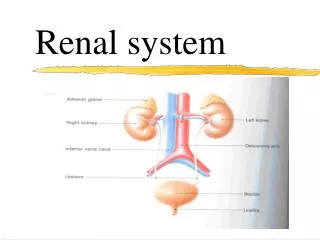
Kidney. Each kidney is enveloped by a dense connective tissue capsule. On the concave side of the kidney, there is an indentation, the hilum, through which the renal artery enters and the renal vein and ureter exit. Deep to the capsule, the parenchyma consists of a cortex and medulla. The medulla is comprised of about 10-18 pyramidal-shaped structures called Pyramids. The base of each pyramid is directed towards the capsule. The apex of each pyramid is called a papilla. The inset is an enlarged view of the papilla. In a longitudinal view, the pyramids appear striated due to the presence of parallel tubules (loops of Henle and collecting ducts) and blood vessels (vasa recta). The cortex is located between the bases of the pyramids and the capsule. Cortical tissue also fills in between the pyramids and is referred to as renal columns. The cortical tissue consists primarily of renal corpuscles and convoluted tubules. Some straight tubules (collecting ducts) project from the pyramids into the cortex. Such an area of the cortex is referred to as pars radiata. The rest of the cortical tissue is referred to as pars convoluta.The central portion of the kidney contains a saccular structure call the renal pelvis (#3). 2-3 tubular structures, major calyces (#2) project from the pelvis. The 2-3 major calyces branch into 10-18 minor calyces (#1). Urine passes from a pyramid into a minor calyx, into a major calyx, and then renal pelvis. The urine then exits the pelvis via the ureter.
Renal Column Capsule Medulla (pyramid) papilla Pars radiata Pars convoluta Cortex Minor calyses Major calyses Renal Pelvis Ureter Renal Sinus PeePee
Kidney. This is a low magnification image of a section through a kidney. The blue line separates the cortex above from the medulla below. The pyramid, containing straight tubules and blood vessels, is cut in cross-section. The cortex contains renal corpuscles (black arrows) and convoluted tubules. A blood vessel (arcuate artery), near the center, is cut in cross-section.
Cortex. These are high magnification images of the cortex of a kidney illustrating two renal corpuscles. These structures consist of a Bowman’s capsule and a tuft of capillaries termed glomeruli (sing= glomerulus). The wall of the Bowman’s capsule is lined by a simple squamous epithelium termed the parietal epithelium (green arrows). The surface of the capillaries is covered by a visceral epithelium. While the cells of the visceral epithelium are classified as a simple squamous epithelium, they are termed podocytes due to their specialized structure. The space located between the glomerulus and the parietal epithelium is termed the urinary space (U).
Renal corpuscle. This is a high magnification image of a renal corpuscle illustrating the parietal epithelium (black arrows) and a glomerulus (glom). Most of the nuclei associated with the glomerulus are those of the podocytes. A urinary space (U) is located between the glomerulus and the parietal epithelium.
Cortical tubules. These are high magnification images of two types of tubules located in the cortex: proximal and distal convoluted tubules. These tubules are lined by a simple cuboidal epithelium. Collecting ducts are not illustrated here. The red arrow is pointing to a proximal convoluted tubule (cut longitudinally) projecting off a renal corpuscle. Urine enters these tubules from the urinary space. The black arrows illustrate proximal convoluted tubules cut in cross-section. These tubules have very irregular lumenal surfaces due to the presence of microvilli. They tend to stain the darkest of the three types of cortical tubules. The green arrows illustrate cross-sections of distal convoluted tubules. These tend to stain lighter than the PCTs and have a smooth lumenal surface.
Arcuate artery. This image illustrates a cross-section through an arcuate artery, which passes along the corticomedullary junction (and parallel to the surface). These arteries are branches of interlobar arteries. Interlobular arteries branch from the arcuate arteries and pass into the cortex along a path indicated by the red arrow. These arteries then give rise to afferent arteries, which supply blood to the glomeruli.
Medulla. These images are cross-sections through a pyramid illustrating three types of medullary tubules. As the PCTs pass into the medulla, they become Thin Loops of Henle (red arrows). These tubules are lined by simple squamous epithelium. The thin loops of Henle are continuous with Thick Loops of Henle (TLH), which pass towards the cortex. These latter tubules, lined by a simple cuboidal epithelium, pass into the cortex as distal convoluted tubules. Structurally, the thick loops of Henle and the DCTs are similar. They are identified by their location. Collecting ducts can be distinguished from thick loops of Henle in that the cells of the collecting ducts tend to bulge toward the lumen and cell-to-cell borders are fairly easy to distinguish. Cell-to-cell borders are difficult to distinguish in the thick loops of Henle.
Medulla. This image is of a cross-section through a pyramid illustrating several thin loops of Henle (red arrows) and some collecting ducts (CD). See previous page for their descriptions.
Papilla. The image on the left is a scanning electron micrograph of a papilla within a minor calyx. Urine passes from the collecting ducts of the papilla (also known as papillary ducts) to the minor calyx. The image on the right is a light microscopic image of a papilla (area within the blue circle in the left image). Collecting ducts (CD) are seen opening into the minor calyx to the left.
Macula Densa. Ascending into the cortex, the DCT approximates its own renal corpuscle. As it does so, there appears on the side of the wall of the DCT facing the renal corpuscle a collection of cells known as the macula densa. These cells are readily identifiable as the nuclei are very close together (red arrows). The macula densa and the J-G cells of the afferent arterioles constitute the juxtaglomerular apparatus
Transitional Epithelium. Transitional epithelium (red circle) lines the caleces, pelvis, ureter, bladder and part of the urethra. It is a stratified epithelium consisting mostly of polygonal cells. The surface cells (green arrows) have a modified surface to withstand the high tonicity of the urine. These cells tend to bulge towards the lumen and are known as “dome” or “balloon” cells.
Ureter. The image on the left is a low magnification image of a ureter. Note that the mucosa (epithelium (red arrows) and underlying lamina propria (black arrows)) contain high rising folds giving it a corrugated appearance. The outer portion of the wall contains smooth muscle (blue arrows). The image on the right is a high magnification of one of the folds. The “dome” cells are not obvious in these sections.
Urinary Bladder. The image on the left is a low magnification of a urinary bladder. The mucosa consists of transitional epithelium (black arrows) and lamina propria (green arrows). The black line indicated the transition between the mucosa and the muscular coat. The muscular coat consists of three layer of smooth muscle: an inner longitudinal layer (above the blue line) a middle circular layer (below the blue line) and an outer longitudinal layer (not shown) The image on the right is a higher magnification illustrating the mucosa.
Think you know your stuff now? Go on and try to get everything right. If you can, you will get 100% on the lab part. If not, maybe medicine isn’t your calling!!!
PCT (note dark staining) DCT (note light staining)
Papilla Pyramid Minor Calyx Minor Calyx
DCT (note smooth lumen) PCT PCT (note irregular lumen)
Renal corpuscle ??? Cortex Arcuate artery ??? Medulla
CD Collecting Duct Thin Loop of Henle Thin Loop of Henle
Collecting Ducts (note the peepee) ? Minor Calyx
Renal corpuscle Glomerulus Bowman’s capsule PeePee space
Smooth muscle: orientation? Longitudinal Lamina propria Transitional epithelium
Macula Densa + JG cells of afferent arterioles = ??? JG Apparatus Glomerulus DCT Macula Densa
Thin Loop of Henle 7. ? TLH 4. ? TLH 1. ? TLH 5. ? TLH 2. ? TLH 6. ? CD 3. ? CD 8. ? TLH Thin Loop of Henle
Type of Tissue? Transitional Epithelium Surface cells (aka Dome Cell, aka Balloon Cell)
PeePee space Hint ?? Glomerulus
Dome (balloon) cells Lamina Propria
Transitional epithelium Lamina propria Layers ?: Epithelium Longitudinal Smooth m. Circular Smooth m.
What tissue is this? Ureter Mucosa Lamina propria Smooth Muscle
If you can get this one, You deserve an award Collagen fibers Elastic fibers Connective tissue cell nucleus Mast cells