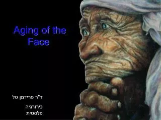
Aging of the Face
480 likes | 856 Vues
Aging of the Face . ד"ר פרידמן טל כירורגיה פלסטית בי"ח אסף- הרופא. Aging of the Face. The process of facial aging represents a combination of gravitational effects and the aging of tissues. Gravity. Affects all tissue layers

Aging of the Face
E N D
Presentation Transcript
Aging of the Face ד"ר פרידמן טל כירורגיה פלסטית בי"ח אסף- הרופא
Aging of the Face The process of facial aging represents a combination of gravitational effects and the aging of tissues.
Gravity • Affects all tissue layers • Results in: Brow ptosis, Hallow infraorbital region, Nasolabial folds, Jowls, Submental skin excess.
Pathogenesis of wrinkles • Aging • Actinic damage • Genetic disorders
Aging A process of atrophy
Epidermis • No change in epidermis thickness • Melanocytes • Langerhans cells • Dermal-epidermal junction
Dermis Components of the dermal connective tissue layer: • Ground substance (Glycosaminoglycan gel + proteoglycans) • Elastic fibers (elastin + microfibrillar components( • Collagen ) Type I:III(
General: 6% for a decade, Connective tissue matrix disorganized, avascular and acellular. Ground substance (GAG) • Elastic fibers number and diameter Collagen :Overall collagen content , III/I , Tensile strength of collagen fibril
Skin appendages • Sebaceous glands in size but sebum production • Pacinian and Meissner’s corpuscles - in number • Apocrine glands - in secretion • Eccrine glands - • Terminal hair follicles -
Effects of Age on Skin • Thinning • Shearing forces • Elasticity • Immunologic changes • Increased susceptibility to UV light and cutaneous malignancies.
Actinic Damage Pathognomonic: Dermal elastosis and epidermal dysplasia.
Epidermis • Increase in thickness • Nuclear atypia of keratinocytes and monocytes
Dermis • Thickened degraded elastic fibers: “Basophilic degeneration”, “elastosis”: Degraded collagen and elastin. • Increase of ground substance. • Decrease of mature collagen (type I).
Inherited Skin Disorders • Rare skin conditions that may present as premature skin laxity, or aging.
Ehlers-Danlos Syndrom (Cutis Hyperelastica) • Histology: Abnormal collagen maturation and tissue fragility, increased capillary fragility. • Genetic defect: Lysyl oxidase • Clinical presentation: Hypermobile joints; Thin, friable, and hyperextensile skin; Subcutaneous hemorrhages; Ability to strech the skin- > Shrink back without wrinkling. Atrophic scars. • Rhytidectomy is not recommended
Cutis Laxa • AD, AR, X-Linked • Inadequacy of elastic fibers through-out the body, especially in the skin, lungs and aorta. • 1' presenting symptom: extreme laxity prematurely aged. • AD: Involve only the dermis. • AR: Generalized abnoralities: Emphysema, pulmonary infection, cor pulmonale, and hernias.
Rhytidectomy is beneficial in the absence of cardiorespiratory dis.
Pseudoxanthoma Elasticum • Mechanically stressed skin ( face and sides of neck and axilla) takes on the texture of plucked chicken skin. • Widespread artheriosclerosis- third decade. • Plastic surgery can be beneficial in the absence of vascular disease.
Progeria (Hutchinson-Gilford Syndrome) • Inheritance: AR • Systemic dis.: Growth retardation, Craniofacial disproportion, Baldness, Pinched nose, Protruding ears, Micrognathia, Artheriosclerosis, Shortened life span. • No role for aesthetic surgery
Werner’s syndrome (Adult progeria) • Inheritance: AR • Scleroderma-like indurated patches of skin, Baldness, Aged facies, hypo-hyper-pigmentation Short suture, high-pitched voice, cataract, mild diabetes mellitus, muscle atrophy, osteoporosis, premature arteriosclerosis, neoplasms. • Elective surgical procedures are contraindicated.
Idiopathic skin laxity • Shelley, 1977, “ wrinkles due to idiopathic loss of mid-dermal elastic tissue” • Patchy areas of mid-dermal elastosis manifesting as localized fine wrinkling, without systemic abnormalities
Histology of the aged skin • Hashimoto, 1974- described 2 types of wrinkles in the skin: • Shallow wrinkle- sun protected area, disappears when the skin is stretched. • Deep wrinkle- sun-exposed skin, that does not disappear on stretching.
Kligman- LM and EM: • Cutaneous wrinkles are mechanically induced grooves of the skin in areas of frequent motion. • Tsuji- Histology of wrinkles: • Deep wrinkle- Greater amount of elastotic swelling on either side of the wrinkle than in the linear depression.
Lapiere- Microanatomy of human skin furrow: • Fascial insertions of cutaneous muscles that create surface grooves on surface animation.
Classification of facial wrinkles, Furrows and Folds • Wrinkles- Intrinsic aging and photoaging. • Clinical location: Cheeks, crow’s feet, perioral. • Tissue location: Cutaneous. • Treatment: Resurfacing.
Folds- The result of overlapping skin caused by genetic laxity, intrinsic aging, loss of tone, bony atrophy, gravity, and consequent sagging. • Clinical location: Lids, nasolabial fold, horisontal neck folds. • Tissue location: Muscular. • Treatment: Rhytidectomy, • Blepharoplasty.
Furrows/ Lines: Repeated facial expressions. Clinical location: Forehead, Glabellar, smile lines. Tissue location: Musculocutaneous. Treatment: Muscle resection, Botulinum toxin, injectable skin filler materials.
Face lifting only addresses the effects of gravity It is not a treatment for fine wrinkles!!!
Stigmata of the aging face • Skin: Intrinsic and extrinsic aging. Subcutaneous: Descent of facial fat- skin layer Fat: Gain or loss Retaining ll.- • Much of this change relates to a loss of support from the retaining ll., accompanied by dermal elastosis, and facial lipodystrophy.
Upper Face • Drooping of the eyebrows. • Hooding of the upper eyelids. • Forehead wrinkling. • Glabellar frown lines. • Prominent lines and laxity of the outer canthus ,onto the temple area. • 40% of face- lifted patients have concomitant forehead lift (Owsley)
Middle and Lower face • Midface: Cutaneous descent superficial to the SMAS. • Lower cheek, and neck: Downward migration of the skin-fat-platysma layer.
Mid face • Yousif- Analized changes in facial soft-tissues occuring with age : 1994,5- Historical photographs and photogrammetry:
NLF- Deepening due to descent and anterior projection of the subcutaneous cheek mass.
Marionette line- a fold extending from the labial commissure to the parasymphyseal area
Skull • Bartlett, Grossman and Whitaker,1992: Analysis of 160 skulls: Reduction of facial height, Modest increase in facial width and depth and generalized coarsening of bony prominences.
Vectors of aging • Alter the position and appearance of key anatomic structures: • The vector of aging of the cheek fat and platysma muscle in the lower face is inferomedial • The vector of the orbicularis oculi is inferolateral
Criteria for a youthful neck • Inferior mandibular border • Subhyoid • Thyroid cartilage • SCM • SM-SM angle- 90 degrees.
The Neck • Excess skin. • Excess submental fat. • Excess supraplatysmal or subplatysmal fat. • Platysmal bands. • Large submaxillary gland. • Micrognatia.
Padgett and Stevenson, 1948- Platysmal bands: • The medial borders of the platysma m. tend to become redundant Platysmal bands. • McKinney, 1995: • The bands are not the medial edge of the muscle, but rather lateral pleats caused by laxity of the muscle . • Pseudoherniation of the submental fat: “Turkey gobbler” deformity.