Download
1 / 21
220 likes | 424 Vues
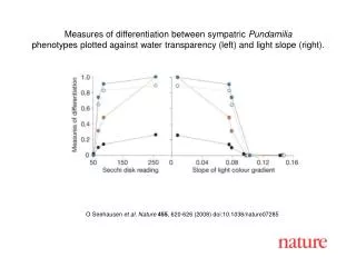
Nanoscale imaging magnetometry with diamond spins under ambient conditions Balasubramanian, G., et al. Nature 455, 648-651 (2008). John Watson 4/15/2009. Outline. Introduction and background Balasubramanian paper Proposed improvements Maze paper Proposed improvements Conclusion.

E N D
Nanoscale imaging magnetometry with diamond spins under ambient conditionsBalasubramanian, G., et al. Nature 455, 648-651 (2008). John Watson 4/15/2009
Outline • Introduction and background • Balasubramanian paper • Proposed improvements • Maze paper • Proposed improvements • Conclusion
Introduction: Why do we care? • Degen, C., Nature Nanotechnology 3, 643-644 (2008).
Introduction: Why do we care? • Room temperature measurements • Biological applications • Single spin detection (electron and neutron) • Quantum experiments – mechanical transport of qubit? • Markers in bio applications
The system • Vacancy produced by electron radiation • Number of vacancies measurable with fluorescence autocorrelation Taylor, J.M., et al., Nature Physics 4, 810-816 (2008).
Method • Optically pump into ms=0 • Split ms=±1 with external B field • Microwave induced dipole transitions • Observe modulation of scattering fluorescence with confocal microscope Right: optically detected magnetic resonance spectra for single nitrogen vacancy
Experiment 1: map probe tip • Look at ESR spectra to determine |B| • Simultaneous AFM to locate vacancy
Resolution • Lock microscope and AFM together • Fixed microwave B • Modulate fluorescence with probe tip • Note sub-wavelength resolution • Dark ring gives maximum resolution – 5 nm
Experiment 2: Vector magnetometer • Excite with microwave field • Scan surface
Results • Shadow due to polarization destruction • 5 mT line represents vacancy resonance • 20 nm line x 25 uT/nm gradient = .5 mT resolution
Proposed improvements • Phase lock with AFM oscillation • Spin echo technique (i.e. ac B field) • Get narrower ESR linewidth • Predicted improvements: 3 uT and sub-nm spatial resolution • Possibly image nuclear spins at 5nm, room temperature
Nanoscale magnetic sensing with an individual electronic spin in diamond. Maze, et al. Nature, 455, 644-647 (2008) • Bulk diamond with vacancy near surface or nanocrystal on substrate • Split degeneracy with DC B field (Helmholtz pair), manipulate with microwave B, spin echo with AC B field, observe ESR spectra with confocal microscopy
Spin echo technique <n> = .03 photons/324 ns • Problem: C13 spins limit coherence time • Solution: apply DC B field to tune frequency to AC field to measure • Measure fluorescence at peaks Nitrogen vacancy spin-echo signal Fluorescence signal
Methods 3.15 kHz • Signal oscillation due to nitrogen spin accumulating phase from AC field • Phase corresponds to spin population difference • Leads to fluorescence variation 4.21 kHz Measured spin echo signal for two operating frequencies. Maximum slope corresponds to maximum sensitivity.
Sensitivity • Expect sensitivity to scale as • Sensitivity optimized for frequencies comparable with spin echo coherence • Photon collection efficiency of .1% limits current sensitivity
Results • Ultrapure bulk sample: 3 nT at kilohertz frequencies with 100 second averaging (roughly 30 nT/Hz-1/2) • Nanocrystal (34 ± 12 nm size): .5 ± .1 μT Hz-1/2 • Stuttgart group (40 nm, single vacancy crystal): .5 mT
Future work • Get samples with fewer 13C isotopes • Improve photon collection with far-field optics or evanescent near-field coupling to waveguides
Conclusion • Proof of principle demonstrated • Much smaller volume than current technology • Can detect ~ 7 electron spins • Significant improvement needed to resolve nuclear spins
References • Nanoscale imaging magnetometry with diamond spins under ambient conditions. Balasubramanian, G., et al. Nature 455, 648-651 (2008). • Nanoscale Magnetometry – Microscopy with Single Spins. Degen, C., Nature Nanotechnology 3, 643-644 (2008). • High-sensitivity diamond magnetometer with nanoscale resolution. Taylor, J.M., et al., Nature Physics 4, 810-816 (2008). • Nanoscale magnetic sensing with an individual electronic spin in diamond. Maze, et al. Nature, 455, 644-647 (2008)