Download
1 / 53
550 likes | 736 Vues
Cardiac imaging in new guidelines and recommendations on… …atrial fibrillation and source of embolism. Paolo Colonna, MD FESC Cardiology Hospital, Policlinico of Bari. august 2010. august 2010. july 2010. Why new recommendations for atrial fibrillation and source of embolism ?.

E N D
Cardiac imaging in new guidelines and recommendations on… …atrial fibrillation and source of embolism Paolo Colonna, MD FESC Cardiology Hospital, Policlinico of Bari
august 2010 july 2010
Why new recommendations for atrial fibrillation and source of embolism ? • Diagnosis of embolism is mainly echo • New technologies (second Ha, deformation, contrast, 3D, etc) + TE echo • Novel and controversial “etiological” therapies for stroke (thrombolysis, closures, ablations, etc) • Changes in stroke population (↑ age and heart failure, ↓rheumatic heart disease)
Origin of Stroke Stroke Registry- St Louis University 26% 29% 3% 4% 37% 1986 1992 Unknown Cardiac Unknown Cardiac 53% Atheroscl. Others Lacunar Lacunar Others Atheroscl. Prior to TEE After TEE Gomez CR, Echocardiography ‘93
ASCO classification. Amarenco P, Cerebrovasc Dis2009;27:502-508 A-S-C-O phenotypes: • A for atherosclerosis, • S for small vessel disease, • C for cardiac source, • O for other cause. Causality levels: • 1: definitely a potential cause of the index stroke • 2: causality uncertain • 3: unlikely a direct cause of the index stroke (but disease is present) • 0: absence of disease
Clinical findings indicating cardioembolic stroke mechanism • Abrupt onset of stroke symptoms, (e.g. AF without preceding TIA / stroke) • Striking stroke severity, old age (NIH-Stroke Scale ≥10; age ≥70 years) • Previous infarctions in various arterial distributions: • Multiplicity in space (= infarct in both anterior and posterior circulation, or bilateral L+R) • Multiplicity in time (= infarct of different age) EAE recommendations, EJE 2010
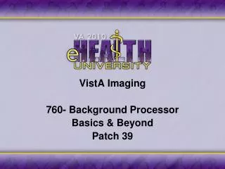
Imaging findings indicating cardioembolic stroke mechanism • Other signs of systemic thromboembolism (e.g. edge-shaped infarctions of kidney or spleen; Osler splits; Blue toe-syndrome) • Territorial distribution of the infarcts • Hyperdense MCA sign (as long as without severe ipsilateral internal carotid stenosis) EAE recommendations, EJE 2010
Territorial distribution Probable Cardioembolic: A) cortex, B) subcortical ‘large lenticulostriate infarct’ Unprobable: C) lacunar infarctions (subcortical) Low flow infarct: interterritorial… D up) subcortical D down) cortical EAE recommendations, EJE 2010
bilateral old infarcts in right middle cerebral artery and left anterior cerebral artery distribution in AF pts hyperdense middle cerebral artery (MCA) sign
(a) Mitral stenosis; (b) Prosthetic heart valve; (c) Myocardial infarction within the past 4 weeks; (d) Mural thrombus in left cavities; (e) Left ventricular aneurysm; (f) History or permanent AF or flutter; (g) Sick sinus syndrome; (h) Dilated cardiomyopathy; (i) Ejection fraction <35%; (j) Endocarditis; (k) Intracardiac mass; (l) PFO plus in situ thrombosis; (m) PFO + PE or DVT preceding the stroke Level of causality 1 (certain)
Level of causality 2 (uncertain) • PFO and ASA; • PFO + DVT or PE (but not preceding the stroke); • Spontaneous echo contrast; • Apical LV akinesia + ↓ ejection fraction (35-50%); • Only suggested: history of myocardial infarction or palpitation and multiple brain infarcts; • Only suggested: abdominal CT/MRI presence of systemic infarction (e.g. kidney, splenic) or lower limb embolism (in addition to the index stroke) • PFO, ASA, valvular strands, mitral annulus calcification, calcified aortic valve, nonapical LV akinesia Level of causality 3 (unlikely)
Multiplane TEE to detect effectiveness of selective pulmonary rt-PA thrombolysis in pulmonary embolism and PFO Colonna, JASE 1997
Paradoxical embolism thrombus in transit through a PFO Srivastava, NEJM 97
Diagnosis and management of entrapped embolus through a PFO Aboyans, EJCTS 98 exhaustive review of the medical literature of this rare finding (43 cases):
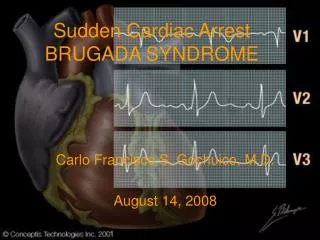
Morphology of PFO in asymptomatic versus symptomatic (stroke or TIA) pts Goel, AJC ‘09
Morphology of PFO in asymptomatic versus symptomatic (stroke or TIA) pts Goel, AJC ‘09 PFOs in pts with cryptogenic CVAs: - larger, - longer tunnels, - more frequently associated with atrial septal aneurysms
Morphology of PFO in asymptomatic versus symptomatic (stroke or TIA) pts Goel, AJC ‘09
Linkage PFO / arterial embolism EAE recommendations, EJE 2010 • Paradoxical embolism through PFO rare, escept in acute pulmonary embolism (↑Right atrium pr) • In the absence of ↑Right atrium pressure, do not suspect causality for PFO, except if: • young age • association ASA + PFO • large right → left shunt • TOE echo + contrasti gold standard for PFO evaluation, but also TT echo (good quality) • Use Valsalva or vigorous cough (TOE and TT ) • Evaluate: color Doppler, n° bubbles, size defect
Echo in AFib / embolic risk EAE recommendations, EJE 2010 Indication of TTecho in AFib for: • diagnosis of cardiac underlying disease (ischemic, valvular, DCM, LV dysfunction) • choose of management and drugs strategy, prior to arrhythmia conversion • indication, guidance and follow up of interventional procedures (ablation, LA appendage closure) Addition of TOecho for: • giudance of TOE/shortened cardioversion • complex cases (embolic recurrences in AC, etc) • additional information on embolic risk(not indicated as a routine exam!)
As alternative to 3 weeks of OAT, the TEE guided cardioversion is recommended to exclude LA or appendage thrombi. Class I LOE B
august 2010 Cardiac imaging and independent risk factors for stroke: • TTE: moderate to severe LV systolic dysfunction • TOE: LA thrombus, complex aortic plaques, spontaneous echo-contrast, and low LAA velocities
Only with TOE Echocardiography in atrial fib: information for clinical decisions EAE recommendations, EJE ‘10 • Thrombi • Spontaneous echocontrast • LA appendage velocities • LV function and thrombi • Patent foramen ovale • Complex aortic plaques Atrio auricular function
TEE correlates of thromboembolism in high-risk patients with nonvalvular AFThe SPAF3 Investigators Committee on EchocardiographyAnn Intern Med 1998 .
Importance of LAA flow as a predictor of thromboembolism in patients with AF Clinical riskfactors Echographicrisk factors Kamp EHJ 99
Prevalence and clinical impact of LA thrombi /echocontrast in AF and low CHADS2 score Kleeman et al. EJE ‘08
Pathophysiologic cascade for stroke in AF pts Clinical risk factors (age, hypertension, etc.) (LV diastolic dysf.) Long lasting AF / Asympt. recurrences Atrio / auricular structural remodeling = Low LAA velocity(LAA dysfunction) Contrast / thrombi in the LAAStroke Khan, Int J Card '03de Luca, Int J Card '05Colonna, JCM '06
Analysis of pts undergoing cardioversion (in ReLY trial) Nagarakanti, Circ 2011
Embolic risk ChadsVasc > 4% 2-3% < 1% > 4 2-3 0-1 Bleedingrisk <1% 3% >10% 0-1 2 >3 Hasbled In doubts… help from echocardiography
for Napoli … in bocca al lupo Echocardiography in stroke and thromboembolism • useful to identify difficult etiologies (masses, endocardites, PFO, thrombi, etc) • study all patients with A Fib for stratification (some of them with TOE) • play “early” to win the championship
Embolic risk stratification of AF pts for the “wise cardiologist” • Calculate %/y embolic risk with CHA2DS2VASc • Calculate %/y bleeding risk with HAS-BLED • In the balance of difficult pts use echo risk factors (atrial appendage, aorta, LV function) • All evaluations more important for new anticoagulants (usage / dosage)
ieri sera… Cagliari Napoli 0-1 Lavezzi al 95’ L’ecocardiografia nello stroke e nel tromboembolismo: • identificare le cause anche quando nascoste • agire presto, ma nei casi difficili non demordere …anche tardi può essere utile per vincere la partita
Imaging findings indicating cardioembolic stroke mechanism • Other signs of systemic thromboembolism (e.g. edge-shaped infarctions of kidney or spleen; Osler splits; Blue toe-syndrome) • Territorial distribution of the infarcts • Hyperdense MCA sign (as long as without severe ipsilateral internal carotid stenosis) • Rapid recanalization of occluded major brain artery (to be evaluated by repetitive neurovascular ultrasound) EAE recommendations, EJE 2010
“At present, closure of patent foramen ovale appears to be reasonable if” : Alp N, Heart 01, mod. • Pt < 60 y with cryptogenetic stroke • Multiple clinical events • Multiple infarcts at CT scan • Valsalva manouver preceding the stroke • Wide PFO (numbers of bubble + dimensions) • Coexistence of atrial septal aneurysm • Deep venous thrombosis Clinical Echo
Stroke mechanisms hypothesis in PFO • Origin from deep vein thrombosis (demonstrated in 5-10%) • Thrombosis in the aneurysm or in the “tunnel” • Increase of atrial arrhythmias • Hypercoagulation state associated
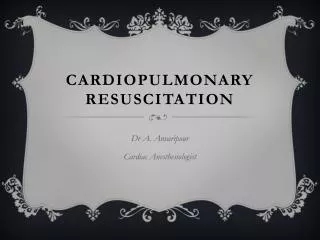
Plaques in thoracic aorta Grade I: normal Grade IV: plaque >4mm Grade II: thickening Grade V: ulcers or mobility Grade III: plaque < 4mm Katz et al JACC ‘92
3 1 2
L’ecocardiografia nel tromboembolismo arterioso: dalle Guidelines dell’EAEPaolo Colonna, MD FESCCardiologia Osp. - Policlinico di Bari
Perché nuove raccomandazioni su ecocardiografia e fonti emboliche ? • Diagnostica embolismo è soprattutto eco • Nuove tecnologie (seconda armonica, deformation, contrasto, 3D, etc.) + ecoTE • Nuove e controverse terapie “eziologiche” per stroke (trombolisi, chiusure, ablazioni, et.) • Cambio popolazione con stroke (↑ età e scompenso, ↓reumatismo)
Eco e diagnosi di endocardite: • Criteri maggiori per diagnosi (3 eco): vegetazioni, ascessi, mobilizzazione di protesi valvolari • Indicato EcoTT precoce in tutti i sospetti clinici • EcoTE se: ecoTT neg. + alto sospetto clinico, protesi valvolari, scarsa qualità ecoTT • Ripetere ecoTT / TE a 7-10 gg se persiste sospetto Eco per predire il rischio di embolizzazione di EI: • Rischio correlato a dimensioni e mobilità: aumentato se vegetazioni grandi (>10 mm), particolarmente se mobili e grandi (>15 mm) • Massimo rischio nei primi giorni dopo inizio antibiotico; decresce dopo 2 settimane EAE recommendations, EJE 2010