
Perimetry visual field
E N D
Presentation Transcript
Perimetry visual field Akram Rismanchian MD Farabi Hospital
The field of vision is defined as the area that is perceived simultaneously by a fixating eye. The limits of the normal field of vision are 60° into the superior field, 75° into the inferior field, 110° temporally, and 60° nasally
THE NORMAL VISUAL FIELD . Traquair, in his classic thesis, described an island of vision in the sea of darkness . The island represents the perceived field of vision, and the sea of darkness is the surrounding areas that are not seen. In the light-adapted state, the island of vision has a steep central peak that corresponds to the fovea, the area of greatest retinal sensitivity.
There are several different packets ofinformation present on the printout , including • Basic identification of patient and test • Indicators of test reliability • Raw unprocessed threshold sensitivity measurements
Deviations of measured sensitivity from age normal ( total deviation ) • Deviation from normal after adjustment for the patients overall sensitivity( pattern deviation ) • Overall indices of normally ( global indices ) • Plain – language analysis ( the glaucoma hemi field test )
Reliability parameters fixation loss false positive response errors false negative response error
Gaze stability • The accuracy of the test result can be strongly affected by the strength or weakness of the patients ability to stay focused on the fixation target during test • The Heiji – Krakau blind spot method can be used to monitor and record a patients fixation loss ( FL ) rate during the test
Perimeters equipped with a gaze monitor show fixation errors on the screen during the test so that perimetrist can make adjustments or instruct the patients when necessary • A tracing appears at the bottom of the printout
False – positive error rates • The perimetric false – false – positive (FP) error rate in the non – SITA strategies is the frequency with which the patient presses during FP catch trials , which are pauses during which no stimulus is presented • The letter X is printed twice next to the FP ratio when it exceeds 33%
The SITA strategies do not make explicit FP catch trials but calculate FP rates from responses at unexpected times during the test m
False – negative error rates • The perimetric false – negative (F N) error rates the frequency with which the patient fails to press the response button when a visible stimulus is presented
For tests run using the full threshold and Fastpac strategies .the false negative rate (FN) is expressed the printout as the ratio of the number of times patients failed to respond to a stimulus 9 db more intense than the previously determined threshold estimate divided by the number of such FN catch trials • As with false – positive responses an XX appears next FN rate on the printout when it exceeds 33%
For tests using the SITA strategies the result is displayed as a percentage instead of ratio • The recorded parameter more closely represents the percentage of time the patient fails respond to visible stimuli because of inconsistency or inattentiveness because the strategy dose not consider any failure to respond during the test that might be caused by disease
Test labeled as having low reliability • Filed were excluded if the fixation loss rate was greater than 20% or if either the false positive rate or the false – positive rate the false – negative rate exceed 33%( in sita stratgy false +ve =8% false – ve =12% )
Raw test results • a) Threshold sensitivity values • Decibel threshold sensitivity values are displayed as a ( sensitivity value table ) , except that the sensitivity value at the fovea is listed over to the side along with the reliability parameters • b) Gray scale
Total deviation ( from normal values for age) • The total deviation display has two parts : a table of numeric values , which represents the actual decibel deviation from age – normal , and below that , a probability plot showing symbols that indicate the statistical significance of each measured deviation
Deviations of less than 5 db may be note worthy near the center of the filed and that even 10 – db deviations near the edge of the visual field may well within the normal range
Pattern deviation ( localized loss ) • The function of the pattern deviation plot is to expose localized defects that may be masked by either a generalized depression or an elevation of the hill of vision • This is accomplished by making an adjustment of the threshold values according to the general height ( GH) of the visual filed
Thus , of the 51 points that are finally considered , the seventh – highest sensitivity value relative to age – normal is taken to represent the overall general height of hill of vision
A key to the probability symbols is shown near the bottom of the printout . • The symbols increase in darkness as the deviation become more significant
Marking a test location with the symbol for p<1% means that fewer than 1% of reliable normal fields in the age –corrected database have sensitivity value that low
Global indices • As many as four global indices provided by the stat Pac analysis including • Mean deviation ( MD) • Pattern standard deviation ( PSD) • Short – term punctuation ( SF ) • Corrected pattern standard deviation ( CPSD)
Mean deviation • The MD index signifies average overall severity of field less • In principle it is the average of all the numbers shown in the total deviation plot
A positive number indicates that the average sensitivity is above - average normal for age . • Whereas a negative number indicates that the average sensitivity is below the average normal value
Pattern standard deviation • Pattern standard deviation ( PSD ) is an index of the roughness of the hill of vision and indicates the degree to which the numbers in the total deviation plot are similar to each other • Specifically the PSD index is the standard deviation around the mean that constitutes the MD index
Short –term fluctuation • The short – term fluctuation ( SF) is in principle the standard deviation of multiple measurement of threshold within a test session • It is an index of interest measurement variability
Corrected pattern standard deviation( CPSD) • The corrected pattern standard deviation (CPSD) index is calculated as an adjustment to the PSD • In an effort to create an index that more purely represent actual unevenness in the amount of filed loss the testing variability, as represent by the SF ,is removed from the PSD to produce the CPSD
Plain language analysis • The Humphery glaucoma hemi filed test ( GHT ) gives a plain language analysis of the visual field test result
In the GHT .five zones in the upper field are compared with five zones in mirror image locations in the lower filed
Outside normal limits Within normal limits Borderline General reduction of sensitivity Abnormally high sensitivity
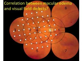
Glaucomatous field deffects 1. Paracentral scotomas 2. An arcuate zone that begins at the blind spot and expands nasally around the point of fixation or there is a depression in the nasal filed of vision , or both
Usually , but not always , the upper portion of the field is more affected than the lower half
3. Hemifield asymmetry and nasal steps The degree of damage at the upper pole of the optic disc usually differs from the degree of damage at the corresponding sector of the lower pole Therefore the threshold visual sensitivities at locations in the upper and lower hemi fields are likely to deviate from normal values unequally
4. Least commonly , localized loss of tissue on the nasal side of the optic disc may produce a “ temporal wedge” A wedge – shaped region of reduced sensitivity with its apex at the physiologic blind spot
DIFFUSE DEPRESSION Diffuse depression of the visual field results from an overall or widespread sinking of the island of vision and may reflect diffuse loss of nerve fibers of the retina. Diffuse depression is a nonspecific sign that can be caused by many etiologies other than glaucoma. By far the most common reason for a diffuse depression is lens opacity. Other factors include other media opacities, miosis, improper refraction, patient fatigue, inattentiveness or inexperience with the examination, ocular anomalies, and age. It is difficult to attribute diffuse depression specifically to a glaucomatous process.
Point of fixation Although a central island of vision typically is preserved until late in the course of glaucomatous optic nerve damage , visual function at the point of fixation may be affected either by diffuse loss of axons ( generalized depression of the field ) or by localized filed defect impinging on the point of fixation )
ضمن عرض پوزش بدلیل حجم بالای LECTUER ادامه اسلایدها امکان پذیر نمیباشد در صورت نیاز به ادامه لطفا به واحد سمعی و بصری مرکز آموزشی درمانی فیض مراجعه و یا با شماره تلفن 03114476010 داخلی 392تماس حاصل نمائید با تشکر