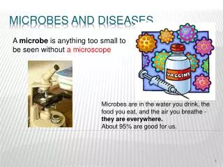
Microbes and Man
Microbes and Man. Infectious diseases - leading cause of death Emerging and re-emerging diseases Development of antibiotic resistance Infections in immunocompromised individuals Importance of the normal flora in human health. Host-Microbe Relationships. Normal flora – transient, permanent

Microbes and Man
E N D
Presentation Transcript
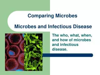
Microbes and Man • Infectious diseases - leading cause of death • Emerging and re-emerging diseases • Development of antibiotic resistance • Infections in immunocompromised individuals • Importance of the normal flora in human health
Host-Microbe Relationships • Normal flora – transient, permanent • Symbiosis (mutualism) – both partners benefit • Parasitic – one benefits, one is harmed (Chlamydia – energy dependencen on host) • Commensal – one benefits, the other is unaffected (anaerobes and aerobes of the GI tract) Relationships can change
The Normal Flora • Play a key role in protecting us from pathogens • Represent the most common causes of bacterial infections in humans • Composition varies with niche and is in constant flux – collectively we carry hundreds of species • Composition is influenced by environmental variables
Environmental variables and Normal Flora • Oxygen concentration varies with niche (air – 21%; mouth – 12%; subgingiva - 1%) anaerobic aerobic microaerophilic • Aerotolerant – Superoxide dismutase, catalase • Temperature, pH, diet, age, hormonal state • Most clinically significant bacteria are aerotolerant
Normal Flora in specific niches (1) • Skin – hostile environment, variable and subject to extreme changes, generally acidic, high in salt, gram + dominate (very few gram -), pores and sweat glands are protected by lysozyme and toxic lipids • Nares – only a few species, usually transient, • Oral cavity – highly variable complex ecosystems, anaerobes dominate (100:1), diet and hygiene influence composition, site of the most prevalent infections of man (dental caries, gingivitis, plaque)
Normal Flora in specific niches (2) • Esophagus – devoid of microflora – mechanical actions make colonization difficult • Stomach – highly acidic, only a few species present, interact tightly with epithelial cells for protection, Helicobacter and Lactobacilli • Small intestine – only a few species, rapid peristalsis, acidic pH, anaerobes
Normal Flora in specific niches (3) • Lg intestine - >400 species, 1011 bacterial cells per gram feces, anaerobes dominate (1000 to 1), E. coli comprises only 1% but is the dominant aerobe • Urogenital tract – only the anterior urethra and vagina are permanently colonized, composition is influenced by hormonal state, lactobacilli are dominant and protect by maintaining low pH
Benefits of the Normal Flora • Food metabolism - proteolytic enzymes • Vitamin Production – produce essential vitamins • Protective effects – compete with foreign organisms for nutrients and receptors, produce antibacterials (Bacteriocins – toxic metabolic by products) • Immune stimulation
Normal flora and Immune Stimulation: Antigen Sampling
Gnotobiotic animals • Animals that are devoid of normal flora • Thin intestinal walls • Under developed Lymphoid tissue • Low antibody titers • Low metabolism rate Provide evidence that normal flora are beneficial
Normal flora and human disease • Leading cause of most bacterial infections in man • Relocation - leads to infection and disease • Streptococcus mutans – dental caries • E. coli and urinary tract infection • Pseudomonas aeruginosa - leading cause of death in cystic fibrosis patients
Microbial Pathogenesis: important terms • Infection • Asymptomatic infection • Pathogenic versus virulent • ID50 and LD50 • toxigenic
Infection: initial steps • Initial interaction and entry – numerous routes, vectors, fomites, direct contact • mucosa - often the initial site of interaction, contains mucin which harbors bacteriostatic agents and provides washing action, sIgA • Goblet cells produce mucin • Adherence and attachment – necessary for the establishment of a productive population
Vector Borne Pathogens
Sources of Infection - Water Cholera Giardia
Adherence and Attachment: a highly specific process Species and tissue tropisms Bordetella pertussis adheres only to ciliated cells
Pili and Fimbriae • - Long, flexible rod-like structures • 100 to 300 per cell • Width 7 nm – diameter 0.2 to 2 um • overcome electrostatic repulsions • Pilin protein – 17 to 25 kDa, form a helical cylindrical structure • tip structure contains adhesins E. coli
Adherence: The Pili – Receptor Interaction The P pili of E. coli binds to a Globobiose Receptor – leads to upper urinary tract infections Receptors in general often contain carbohydrates or glycolipids
Variable Adherence Mechanisms of E. coli • E. coli virotypes – ETEC, EHEC, EPEC • Enterotoxigenic E. coli – adhere to ciliated cells (sm intestine), cause no direct damage. Secrete toxins that diffuse and interact w/ host cells • Enteropathogenic E. coli – adhere tighly to non-ciliated cells, induce changes in host cell cytoskeleton, leads to effacing and tissue damage
Adherence of Enterotoxigenic E. coli Loose interaction - sufficient to allow toxin to diffuse to the target cells
Enteropathogenic E. coli: Attachment and Effacing Tight interaction, effacing occurs, inflammation and severe tissue destruction
Attaching and Effacing by E. coli E. coli EPEC – induce rearrangement of actin filaments in the host cell. The process is mediated by the eae genes (E. coli attachment and effacing Pedestal formation
Attachment of Vibrio cholerae to Host Cells Vibrios adhering to intestinal epithelial cells Vibrios adhering to M cells – induction of host cell rearrangement
Afimbrial Adhesins Used by many bacteria, particularly Gram (+) Individual proteins or clusters of protein on the cell suface – mediate a tight interaction with host cells Afimbrial adhesins on the cell surface bacteria
Biofilm Formation Staphyloccocus epidermidis in the early stages of biofilm formation Biofilms formed by normal flora may prevent the binding of pathogens (competition). Pathogens also form biofilms (dental plaque, catheters, implants Biofilms protect against antibiotics, antibody, complement and phagocytosis
Bacterial Invasion • Why invade? • obligate, facultative, transient, permanent • nutrient rich environment • protective from immune system • cooperative process involving cytoskeleton rearrangement in the host Listeria – enters and moves through cell using actin rearrangement
Invasion by Salmonella typhimirium Bacteria interact with host cells Induce rearrangements Internalize Replicate in vacuoles Lyse cell and escape
Capsules and Pathogenesis • Loose unstructured network of polysaccharides • Protects against phagocytosis • Aids in adherence • Inhibits complement activation through binding of factor H- Klebsiella
Capsules and Pathogenesis • Layer of exopolysaccharides and protein • Relatively water insoluble • Can function in adherence • Protect against phagocytosis • Bind factor H to circumvent complement by preventing C3 convertase formation • Can be highly immunogenic (employed in vaccine development – PRP capsular vaccine for Haemophilus influenza type b) Klebsiella pneumonia
IgG and IgA Proteases • Haemophilus influenzae; S. pneumoniae • Targets vary; IgG, IgA • IgA proteases facilitate mucosal survival • IgA is a natural defense of the mucosa Antiphagocytic Factors • Staphylococcus aureus - Protein A (binds Fc region of IgG) • Streptococcus pyogenes - M Protein (binds factor H) • -chemical modification of capsule and LPS (sialylation)
Role of Factor H in the Alternative Pathway of the Complement Cascade Factor H binding is an immune evasion mechanism employed several bacteria, viruses and fungi
Factor H Binding to Surface Proteins: Evasion of complement attack 1 2 3 4 5 6 7 8 9 10 11 12 13 14 • Proteins from Lyme disease isolates were fractionated by SDS-PAGE electrophoresis • Proteins were transferred onto membranes for Western blotting • The membranes were incubated with factor H • Anti-factor H antisera was used to detect factor H- borrelia protein complexes
Lyme disease: VlsE, OspE and Antigenic Variation • Plasmid carried genes • Surface exposed lipoproteins • Highly immunogenic • VlsE: 1 functional VlsE gene and 16 partial gene cassettes - segmental gene conversions alter sequence of the expressed gene • OspE: homologous recombination among related genes
Antigenic Variation in Lyme Disease: A Chronic Infection Segmental gene conversion - unidirectional Expression locus
OspF SNP Analysis: BBO39 is Stable During Infection in Mice 17 17 14 24 14 24 pc 15 pc 15 7 9 9 7 BBO39 (an ospF gene) vlsE
Variable Regions 1-5 of VlsE Variants VR-1 VR-2 VlsEpc ANAGAAKAADKDSVK GGSEKLK-VAAAKGENNKK VlsEc17 .-DN.......E... ............T..-.E. VlsEc53 –-DN..........T ............T..…. VlsEc53e13 –-DND......A..T R.............-G.E. VlsEc53e72 –-DND..V...E..T R.............-G.E. VR-3 VR-4 VR-5 VlsEpc VGDA-A KAAG–-AAEQDGKKPAEAK DA-DGADF-KDE VlsEc17 ...... ...........E........ ..E...E..... VlsEc53 A..LLC .....EA.GD.E....ED.. .KDGD.E.G-.G VlsEc53e13 A..DN. T............E..GD.. N.D.....N.EG VlsEc53e72 A..DN. T............E..GD.. N.D.....N.EG
Exotoxins and Pathogenesis • Soluble, secreted, heat labile molecules • Toxin nomenclature – based on species of origin, activity, or target substrate • Role in disease – some cause disease in the absence of the bacteria while others do not • ADP-ribosylating toxins – diverse group of toxins with separable activity and binding domains
ADP Ribosylating Toxins: Structure and Processing Simple toxins – A and B domains are encoded by the same gene Compound toxins – A and B are encoded by different genes Processing: for secretion and maximal activity, all AB toxins undergo processing. A and B components remain linked by a disulfide bond
ADP Ribosylating Toxins ADP Ribose moiety is derived from NADH All have AB structure and require processing In spite of sequence variation in this class of toxins they all catalyze a similar rection but target different molecules
Cholera Toxin: Expression and Structure • Vibrio cholera – g(-), not all strains express toxin • Toxin is phage encoded (toxigenic strains) • Operon: ctxA and ctxB • 5 B units and 1 A • Expression and ribosomal binding sites
Cholera Toxin: Secretion -components are synthesized in the cytoplasm - transported to the periplasm and assembled -secreted and the A subunit is cleaved into A1 and A2
Internalization of Cholera Toxin Receptor: GM1 ganglioside located on cells of the small intestine Only the A1 subunit is released into the cytoplasm
Cholera Toxin: Mechanism of Action • A1 ADP ribosylates an Arg of the Gs protein locking it in the “on” state • Gs positively regulates adenyl cyclase • This leads to uncontrolled cAMP production • Na and K transport systems are disrupted – leads to outflow of Cl and H2O into the intestinal lumen • Severe diarrhea (20 liters day-1) and electrolyte loss • Hypotension and vascular collapse
Same Enzymatic Activity – Different Targets - Diphtheria Toxin: an ADP Ribosylating Toxin
Pore Forming and Membrane Disrupting Toxins Pore forming: Aggregates in membrane forming a pore. Ex; Listeriolysin O (escape from phagosomes) Membrane disrupting: cleaves the polar head group from phospholipids destabilizing the membrane Ex; Listeria - phospholipase C
Listeriolysin O and Listeriosis • Bacteria adhere to intestinal mucosa • They produce internalin - stimulates phagocytosis • Phagosomal escape – phagosome acidifies before lysosomal fusion, LLO becomes active at pH 5.5, forms pores in the phagosome, bacteria escape, LLO is inactive in the cytoplasm