Acute Abdomen
640 likes | 733 Vues
Learn about acute abdominal pain, its importance, diagnostic process, patient examination, and symptom evaluation. Understand types of pain, related symptoms, and consider scenarios like in children and pregnant women.

Acute Abdomen
E N D
Presentation Transcript
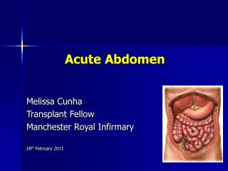
Acute Abdomen Nurhayat Usman, dr Sp.B-KBD
Acute Abdomen (acute abdominal pain) • “Condition which requires immediate treatment” (FD Moore, 1977): Surgery? When to perform? • (Buku Ajar Ilmu Bedah, 1997): “Clinical condition which arises from acute critical condition in the abdominal cavity, and usually manifests as pain. • Acute abdominal pain: Chief complaint: acute pain (Nyhus, Vitello, Condon, 1995)
Why is it important ? • Patient with acute abdomen: • Sudden onset • Unknown etiology (not clear) • Need immediate diagnosis & treatment • Prevent morbidity & mortality
Morbidity & Mortality Obstruction Fluid imbalance Perforated viscus Peritonitis Infection Sepsis Shock Bleeding Hypovolemic Shock Ischaemia Perforation Peritonitis Death
Acute abdominal pain • Most can be diagnosed clinically • Require accurate and focused history taking • Need meticulous & rationale physical examination • Appropriate special investigations
TheDiagnosticProcess HISTORY Patient perception of symptoms Patient description of symptoms Physician perception Physician interpretation of symptoms LABORATORY SYNTHESIS PHYSICALFINDINGS RECORDING EXAM DECISION
History Taking • 60 - 80% of accurate diagnosis arises from good & meticulous history taking • Physical diagnosis confirms accurate diagnosis • 10 - 15% of accurate diagnosis arise from laboratory & radiological examinations
History Taking • May confirm : • Suspected diagnosis • Possible etiology • Disease stages/ complications • Differential diagnosis
Patient Identity • Ask the patient politely concerning his/her : name age • Record the gender : • Male • Female • Ask the marital status of the patient (especially for female)
Acute abdominal pain in specific groups • In children • Acute appendicitis • In the elderly • Perforated tumors • Bowel obstruction due to tumors • During pregnancy • Complicated Ectopic pregnancy
Chief complaint: Ask the patient regarding why the patient comes to you. Onset PAIN Site at onset Radiation Site at present Type Progression Severity Duration Aggravating /relieving factors
Upper abdominal pain • Peptic or gastric ulcer • Acute Cholecystitis, Acute Cholangitis • Pancreatitis • Early Appendicitis • Hepatitis or liver abscess • Extra abdominal: • Inferior Pleuritis, lobar pneumonia, pneumothorax • Pericarditis, Myocardial infarction, angina • Pyelonephritis, renal colic
Central abdominal pain • Early appendicitis • Bowel obstruction, strangulated • Pancreatitis • Gastroenteritis • Mesenterial Emboli /Thrombosis • Dissecting aortic aneurism • Mesenteric adenitis • Early sigmoid diverticulitis
Lower abdominal pain • Colonic Gangrene/Obstruction • Appendicitis • Mesenteric adenitis • Diverticulitis • Ruptured tubo-ovarial abscess • Tuboovarial Torsion • Ectopic gestation
Onset of pain • Sudden onset
Onset of pain • Gradual pain
Type of pain Visceral pain & Parietal pain
Type and severity of pain • A. Toothache • C. Colicky pain of inflammed hollow organs A C
Type and severity of pain • Intermittent colicky pain of obstructed hollow organ at early stage.
Type and severity of pain • Progressive & Continous colicky pain due to strangulated bowel obstruction (ischemic stage)
Other related symptoms:Ask the patient concerning related/concomitant symptoms of • Gastro-intestinal function: • Nausea • Vomiting • Loss of appetite • Faintness • Previous indigestion (habitual)
Other related symptoms: • Jaundice • Bowel habit: • Constipation? • Diarrhoea? • Colour of the stool? • Presence or absence of blood and mucus (slime)
Other related symptoms: • Urinary function: • Micturition: amount of urine, lower abdominal discomfort, colour of urine • Gynaecological function (female) • Menstrual function • Delayed or miss period • Abnormal bleeding or discharge (colour, quantity)
Previous history • Similar pain • Abdominal surgery • Major illness: incl. fever, abdominal injury. • Drugs • Allergies
PHYSICAL EXAMINATION • Preparation • Check all the equipment required and have a good light: • Examination couch • Stethoscope • Explain the procedure and its goals to the patient. • Wash your hands with antiseptic soap. • Dry and warm your hands with tissues.
Implementation: • A General Examination • General appearance:Consciousness • Mood: Distressed? Anxious? • Immobile • Move cautiously • Colour: Pallor? Flushing? Jaundice? Cyanosis?
Implementation: • Examine the vital signs: • Temperature • Pulse rate • Blood Pressure • Respiratory rate
Implementation: • Perform other systems examination, including cardio-pulmonary system. • Ask the patient politely to expose his/her abdomen.
Abdominal Examination: Inspection • Inspect the movement: • Respiratory movement • Visible bowel peristaltics • Is there any scars on the skin of the abdomen? • Is there any abdominal distention? • Flatus ? , Fluid ? , Fetus?
Abdominal Examination: Inspection • Is there any rashes and discolouration? • Cullen’s sign • Gray Turner’s sign • Ecchymosis of the abdominal wall • Is there any masses: • Tumors? • Hernial sites? • Masses with pulsation?
Gray-Turner Sign Cullen Sign
Abdominal Examination: Palpation • Ask the patient to locate the site of maximum pain with the tip of a finger. • Using the palmar surface of your fingers, gently palpate the abdomen, starting from a site farthest from the area of maximum pain, move gradually towards it.
While palpating, look to the face expression of the patient, and look for any signs of : • Tenderness • Rebound tenderness • Muscle guarding • Rigidity • Murphy’s sign
While palpating, look to the face expression of the patient, and look for any signs of : • Swelling or masses • Rovsing’s sign • Expansile pulsation • Hernial orifices • Scrotum in male
Specific signs • Rovsing’s sign • Obturator sign • Psoas sign
Abdominal Examination :Percussion • Place the palmar aspect of your left hand on the abdomen, and gently percus its dorsal aspect with the tip of the middle finger of the right hand, moving all around the abdominal region: • Is it tymphanitic? • Is it dull ? • Is there any shifting dullness? • Site of liver dullness ? and is it disappeared ?
Auscultation • Using stethoscope, and place it gently on the abdomen, listen to the bowel sounds and bruit at least for one minute: • Absent? • High pitched and hyperactive? • Metallic sound? • Vascular bruit?
Digital Rectal Examination • Put on surgical hand gloves and ask the patient to expose his/her buttock and anus, and place the patient in lithotomy position.Apply lubricating jelly on to the right index finger.
Digital Rectal Examination • Gently insert your right index finger into the anus, move toward the anal canal slowly, and evaluate the followings: • Anal margin: piles? • Mucosal surface of the anal canal and the ampulla (collaps?) • Sites of any pain elicited • Masses or swelling: consistency, location, surface, fixity to the surroundings. • Bowel contents: consistency of faeces? Mucus? Blood?
Perform bimanual palpation in female patient to examine the uterus, pelvic cavity and adnexa. • Write up • Write up all significant findings in the medical record. Conclude your diagnosis and differential diagnosis, and order any necessary special investigations
Extraperitonealcauses of acute abdomen • Cardiothorax • Urology • Vascular • E.t.c
Degree of peritoneal irritation (Lowenfels, 1975) Bowel bontent Gastric juice Pancreatic juice pus Urine blood bile Mild Severe
Signs of Intrabdominal Sepsis • Fever, nausea, vomiting, tachicardia, tachipneu • Abdominal pain • Peritoneal signs • Signs of dehydration • Leucositosis • Shock, Multiple organ failure
Tips • > 6 hours: surgically related diseases !!! • Limited movement: peritonitis / ischaemia • Persistent pain on morphine : ischaemia • Sense of Crisis • Repeated exams : Very important