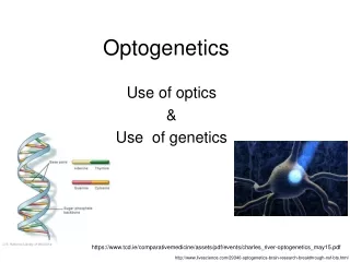
Optogenetics: Evolution and Innovative Applications
Explore the history and advancements in optogenetics, from the discovery of opsins in the 1970s to current research using light-activated compounds for neural modulation and cell activation. Learn about key breakthroughs and the collaboration between scientists driving this field forward.

Optogenetics: Evolution and Innovative Applications
E N D
Presentation Transcript
OPTOGENETICS andCaged compounds Saad Abbasi, Lindsey Biggs and Brennan Paedae
History of the development of optogenetics Paper published by Chow et al, (2010) using archaerhodopsin which completely shuts down the cell. Collaboration between Nagel, and Deisseroth (and Boyden) Three-gene phototransduction cascade used to activate cells Halorhodopsin used for neural silencing Discovery and study of opsins 1970’s 1999 Paper on channelrhodopsin published by Georg Nagel et al. (2003) Use of nanoparticles and magnetic pulses to activate specific cell types without such invasive measures (Palle Lab?) Papers published using OptoXR, light activated GPCR’s which modulate intracellular signalling (Aiden,2009) and use in live primates (Han et al. 2009) First published paper using optogenetics (Boyden et al., 2005) on cultured hippocampal neurons, followed by papers from several other labs Halorhodopsin and neural chloride levels Photo: Halobacterium salinarum http://www.biochem.mpg.de/en/eg/oesterhelt/web_page_list/Org_Hasal/index.html
1970-1980’sDiscovery and study of opsins • Halobacteriumsalinarum: • Motile organisms • Can live with light as the only energy source (Bacteriorhodopsin) • 4 retinal proteins: • Bacteriorhodopsin: light driven proton pump that converts light to energy source [discovered in early 1970’s (Boyden, 2011)] • Halorhodopsin: chloride pump that maintains salt concentration [discovered in late 1970’s (Boyden, 2011)] • Sensory rhodopsin 1:phototactic response to orange light [discovered in 1980’s (Boyden, 2011)] • Sensory rhodopsin 2: phototactic response to blue light. • BUT….. The organisms expressing these rhodopsins function in environments with high salt concentrations, so there was little optimism for function in neural tissue. http://www.mpi-magdeburg.mpg.de/research/groups/mna/smallnet.html?pp=1 http://www.biochem.mpg.de/en/eg/oesterhelt/web_page_list/Org_Hasal/index.html
1999Halorhopsin and neural chloride levels • Okuno et al. re-opened the possibility of using rhodopsins in neural tissue with his 1999 paper. • Compared to H. salinarum, rhodopsins from Natronomona pharaonis functioned best at chloride concentrations that are similar to concentrations seen in neural tissue. • BUT….. It was still unknown whether these rhodopsins could be expressed and functional in neural tissue.
History of the development of optogenetics Paper published by Chow et al, (2010) using archaerhodopsin which completely shuts down the cell. Collaboration between Nagel, and Deisseroth (and Boyden) Three-gene phototransduction cascade used to activate cells Halorhodopsin used for neural silencing Discovery and study of opsins 1970’s 1999 2002 Paper on channelrhodopsin published by Georg Nagel et al. (2003) Use of nanoparticles and magnetic pulses to activate specific cell types without such invasive measures (Palle Lab?) Papers published using OptoXR, light activated GPCR’s which modulate intracellular signalling (Aiden,2009) and use in live primates (Han et al. 2009) First published paper using optogenetics (Boyden et al., 2005) on cultured hippocampal neurons, followed by papers from several other labs Halorhodopsin and neural chloride levels Photo: chARGe in cultured hippocampal neuron (GFP tagged) (Zemelman, 2002)
2002 chARGe may be the answer! • GeroMiesenbrook and colleagues created a three-gene Drosophiliaphototransduction cascade that could be expressed in cultured hippocampal neurons. When exposed to light, cells expressing chARGe were more active. (Zemelman, 2002). • chARGe= Arrestin-2 rhodopsin coupled to alpha subunit of g-protein. • However, the activation of neurons was not instantaneous, but took several seconds. • A temporally precise method of activation was still necessary.
History of the development of optogenetics Paper published by Chow et al, (2010) using archaerhodopsin which completely shuts down the cell. Collaboration between Nagel, and Deisseroth (and Boyden) Three-gene phototransduction cascade used to activate cells Halorhodopsin used for neural silencing Discovery and study of opsins 1970’s 1999 2002 2003 2004 Let there be light! Channelrhopsin 2 is light sensitive. Use of nanoparticles and magnetic pulses to activate specific cell types without such invasive measures (Palle Lab?) Papers published using OptoXR, light activated GPCR’s which modulate intracellular signalling (Aiden,2009) and use in live primates (Han et al. 2009) First published paper using optogenetics (Boyden et al., 2005) on cultured hippocampal neurons, followed by papers from several other labs Halorhodopsin and neural chloride levels Photo: ChR2 conjugated to RFP http://en.wikipedia.org/wiki/Channelrhodopsin
2003 and 2004Channelrhodopsin-2 • Chlamydomonareinhardtii use channelrhodopsin-2 (ChR2) to drive phototaxis (Sineshchekov et al. 2002). • ChR2 is a light gated cation channel which produces movement in C. reinhardtii. • Nagel and colleaques used ChR2 in oocytes and HEK cells to show that it could be used to depolarize cells via illumination (Nagel et al. 2003). • In 2004, a collaboration between Georg Nagel, Karl Deisseroth (and Edward Boyden) began. • Nagel had since discovered that not much all-trans retinal needed to be added to the cultures for ChR2 function. Boyden, 2011
History of the development of optogenetics Paper published by Chow et al, (2010) using archaerhodopsin which completely shuts down the cell. Collaboration between Nagel, and Deisseroth (and Boyden) Three-gene phototransduction cascade used to activate cells Halorhodopsin used for neural silencing Discovery and study of opsins 1970’s 1999 2002 2003 2004 2005 Let there be light! Channelrhopsin 2 is light sensitive. First published paper using optogenetics (Boyden et al., 2005). Use of nanoparticles and magnetic pulses to activate specific cell types without such invasive measures (Palle Lab?) Papers published using OptoXR, light activated GPCR’s which modulate intracellular signalling (Aiden,2009) and use in live primates (Han et al. 2009) Halorhodopsin and neural chloride levels Photo: ChR2 response to light (cultured hippocampal neuron) (Boyden, 2011 from Boyden et al. 2005 paper)
2005First published paper using optogenetics • In 2005, Edward Boyden, Karl Deisseroth and colleagues published the first paper using optogentics in cultured mammalian hippocampal neurons. • ChR2 was expressed, and functional in neurons. • Current produced by ChR2 activation was enough to produce action potentials. • ChR2 had a low rate of inactivation and quick recovery time. • Several other labs published papers using similar techniques soon after. These methods had been on the minds of many research groups! • Yawo Lab – 11/05- intact mammal brain circuits • Herlitze and Landmesser labs-11/05 chick spinal cord • Nagel and Gottschalk labs-12/05; behaving worm • Pan Lab- 4/06; retina (Boyden, et al. 2005)
History of the development of optogenetics Paper published by Chow et al, (2010) using archaerhodopsin which completely shuts down the cell. Collaboration between Nagel, and Deisseroth (and Boyden) Three-gene phototransduction cascade used to activate cells Halorhodopsin used for neural silencing Discovery and study of opsins 1970’s 1999 2002 2003 2004 2005 2007 Let there be light! Channelrhopsin 2 is light sensitive. First published paper using optogenetics (Boyden et al., 2005). Use of nanoparticles and magnetic pulses to activate specific cell types without such invasive measures (Palle Lab?) Papers published using OptoXR, light activated GPCR’s which modulate intracellular signalling (Aiden,2009) and use in live primates (Han et al. 2009) Halorhodopsin and neural chloride levels Photo: Halorhodpsin Lief et al., 2011
2007N. pharaonisHalorhodpsin • March 2007-Xue Han and Boyden published data showing that N. Pharaonishalorhodopsin could be used for neural silencing. (Cl- channel) • Few weeks later, Karl et al published a paper showing the same conclusion and that it could be used to modify behavior in C. elegans. • BUT…..halorhodopsin had low magnitude currents, would get stuck in inactivation phase after long stimulation, and had a slow recovery period. (Boyden, 2011)
History of the development of optogenetics Paper published by Chow et al, (2010) using archaerhodopsin which completely shuts down the cell. Collaboration between Nagel, and Deisseroth (and Boyden) Three-gene phototransduction cascade used to activate cells Halorhodopsin used for neural silencing Discovery and study of opsins 1970’s 1999 2002 2003 2004 2005 2007 2009 Let there be light! Channelrhopsin 2 is light sensitive. Papers published on optogenetics in primates. First published paper using optogenetics (Boyden et al., 2005). Use of nanoparticles and magnetic pulses to activate specific cell types without such invasive measures (Palle Lab?) Halorhodopsin and neural chloride levels
2009Optogenetics in primates • Boyden and Desimone published research on primate brains, suggesting these methods could someday be used for clinical purposes. • Conclusions: • ChR2 can be expressed in maquaqe monkeys to modulate activity in specific subsets of neurons, without inducing neuron death and immune responses.
History of the development of optogenetics Collaboration between Nagel, and Deisseroth (and Boyden) Three-gene phototransduction cascade used to activate cells Archaerhodopsin completely silences neurons. Halorhodopsin used for neural silencing Discovery and study of opsins 1970’s 1999 2002 2003 2004 2005 2007 2009 2010 Let there be light! Channelrhopsin 2 is light sensitive. Papers published on optogenetics in primates. First published paper using optogenetics (Boyden et al., 2005). Use of nanoparticles and magnetic pulses to activate specific cell types without such invasive measures (Palle Lab?) Halorhodopsin and neural chloride levels
2010Arechearhodopsin • The solution to the limitations of halorhodopsin: Archearhodopsin • The paper published in Jan 2010 by Chow and Boyden showed that archearhodopsin: • Could completely shut down the cell. • Had rapid recovery after long stimulation • hyperpolarize the cell by pumping protons out of the cell (Chow et al. 2010).
History of the development of optogenetics Collaboration between Nagel, and Deisseroth (and Boyden) Three-gene phototransduction cascade used to activate cells Archaerhodopsin completely silences neurons. Halorhodopsin used for neural silencing Discovery and study of opsins 1970’s 1999 2002 2003 2004 2005 2007 2009 2010 2011 Let there be light! Channelrhopsin 2 is light sensitive. Papers published on optogenetics in primates. First published paper using optogenetics (Boyden et al., 2005). Use of nanoparticles and magnetic pulses to activate specific cell types without such invasive measures Halorhodopsin and neural chloride levels Photo:Arnd Pralle, physics prof. at Univ of Buffalo. Research on magnetic nanoparticles
Parkinson’s Disease • Degenerative disorder of the CNS. • Most cases occur after the age of 50 • Causes: • Death of cells in the Substantianigra which produce dopamine. • Cause of cell loss is unknown, but is genetic is some cases. • Symptoms: • Movement-related: shaking (tremor), rigidity, slow movements, difficulty walking and with gait postural instability. • Cognitive and behavioral problems in more advanced stages: dementia, sensory, sleep and emotional problems. • Diagnosis is usually based on symptoms, with neuroimaging used for confirmation. • Treatments: • L-Dopa (which can cross the BBB) and other dopamine agonists. • With the loss of DA producing neurons, these treatments become ineffective and can cause diskinesia (involuntary writhing movements) • Deep-brain stimulation and lesion surgery are used as a last resort. www.wikipedia.org
Basal ganglia circuitry Substantia nigra pars reticulata Substantia nigra pars reticulata Substantia nigra pars reticulata http://www.ncbi.nlm.nih.gov/books/NBK10847/
D1-Cre mice expressed ChR2-YFP in striatum and fibers projecting through globuspallidus to SNr. • D2-Cre mice, ChR2-YFP cells bodies were seen in striatum, projecting to globuspallidus. • ChR2 was expressed in medium spiny neurons (DARPP-32 MSN marker) Supp. Fig. 1
Whole cell slice electrophysiology • Whole cell slice eletrophysiology was used to verify ChR2 expression in D1 and D2 specific neurons. • Current-firing relationship for direct and indirect pathways were consistent with previous data (a,b) • 470 nm illumination of the ChR2 expressing neurosn produced light-evoked inward current and increased spiking.
Silicon probe with integrated laser-couple optical fiber. In vivo laser stimulation and recording
Behavioral data • Activation of direct pathway (D1) increased percent of time in ambulation, and decreased freezing and fine motor movements. • Activation of indirect pathway (D2) decreased ambulation and fine movements and increased the time spent freezing. Causal relationship between direct pathway in increasing motor behavior and between indirect pathway and increased freezing responses.
Bilateral 6-OHDA injections caused loss of dopaminergicinnervation in dorsomedial striatum and Parkinsonian-like motor deficits. • Activation of the direct pathway completely restored pre-lesion motor behaviors. • Decreased freezing • Increased locomotor activity.
Conclusions: • This study provides evidence in a behaving animal that activation of the direct and indirect pathways are modulating motor activity as previously suggested. • This technique offers temporally precise activation of the circuitry (compared to pharmacological blockade, lesions, or transgenic mice). • This technique also allows for a quick return to baseline firing rates and activity. • Activation of the direct pathway in basal ganglia can ameliorate motor deficits caused by loss of striatal neurons (which are modulated by DA release from substantia nigra.)
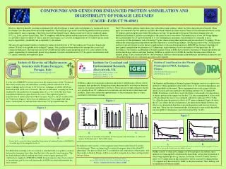
Halorhopsin from H. salinarum functions best at high chloride levels. (Left) • Halorhopsin from N. pharaonis functions best at lower chloride concentrations, which are similar to neural tissue. • (Okuno et al., 1999)
http://www.fmls-institute.de/index.php?id=neurobiochemistry http://blogs.physicstoday.org/industry09/ https://encrypted-tbn0.google.com/images?q=tbn:ANd9GcQL5mvVzT07u1TnAgnMC7QStw5V2BJ3-5ytYFM5lVolKD34QkoEKQ OPTOGENETICS http://www.aan.com/elibrary/neurologytoday/?event=home.showArticle&id=ovid.com:/bib/ovftdb/00132985-201107070-00006 http://czechfood.blogspot.com/2011/07/optogenetics-optogenetica.html Lindsey Biggs and Brennan Paedae http://www.stanford.edu/~shenoy/GroupResearchPublications.htm https://encrypted-tbn1.google.com/images?q=tbn:ANd9GcQVAQDbKMATn9SkXr45jkleAr9O9HqOum4wjihhLB4161OkhNO68w http://www.nytimes.com/2011/05/17/science/17optics.html?_r=1