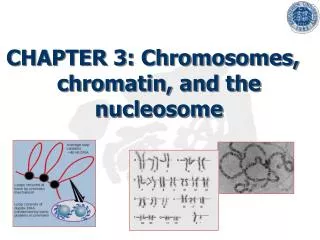
Nucleosome structure
Nucleosome structure. Histones. Most of the protein in eukaryotic chromatin consists of histones , of which there are five families, or classes: H2A, H2B, H3 and H4, known as the core histones ,

Nucleosome structure
E N D
Presentation Transcript
Histones • Most of the protein in eukaryotic chromatin consists of histones, of which there are five families, or classes: H2A, H2B, H3 and H4, known as the core histones, • and H1. The core histones are small proteins, with masses between 10 and 20 kDa, and H1 histones are a little larger at around 23 kDa. • All histone proteins have a large positive charge; between 20 and 30% of their sequences consist of the basic amino acids, lysine and arginine This means that histones will bind very strongly to the negatively charged DNA in forming chromatin.
Histones • Members of the same histone class are very highly conserved between relatively unrelated species, for example between plants and animals, which testifies to their crucial role in chromatin. that all histones are ultimately evolutionarily related • H1 histones are somewhat distinct from the other histone classes in a number of ways; in addition to their larger size, there is more variation in H1 sequences both between and within species than in the other classes.
Nucleosome Stucture • Treatment of chromatin with micrococcal nuclease, an endonuclease which cleaves double-stranded DNA, led to the isolation of DNA fragments with discrete sizes, in multiples of approximately 200 bp. • It was discovered that each 200 bp fragment is associated with an octamer • core of histone proteins, (H2A)2(H2B)2(H3)2(H4)2, which is why these are designated the core histones, and more loosely with one molecule of H1. • The proteins protect the DNA from the action of micrococcal nuclease.
Nucleosome Stucture • prolonged digestion with nuclease leads to the loss of H1 and yields a very resistant structure consisting of 146 bp of DNA associated very tightly with the histone octamer. This structure is known as the nucleosome core, and is structurally very similar whatever the source of the chromatin • The histone octamer forms a wedge-shaped disk, around which the 146 bp of DNA is wrapped in 1.8 turns in a left-handed direction.
Nucleosome Stucture • One molecule of histone H1 binds to the nucleosome, and acts to stabilize the point at which the DNA enters and leaves the nucleosome core • In the presence of H1, a further 20 bp of DNA is protected from nuclease digestion, making 166 bp in all, corresponding to two full turns around the histone octamer. A nucleosome core plus H1 is known as a chromatosome. • In some cell types, it may be replaced by an extreme variant called histone H5, which binds chromatin particularly tightly, and is associated with DNA which is not undergoing transcription
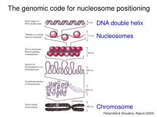
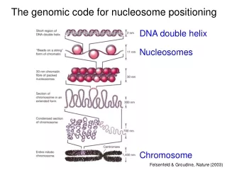
The 30 nm fiber • The presence of histone H1 increases the organization of the ‘beads on a string’ to show a zig-zag structure in electron micrographs. • With a change in the salt concentration, further organization of the nucleosomes into a fiber of 30 nm diameter takes place. • Detailed studies of this process have suggested that the nucleosomes are wound into a higher order left-handed helix, dubbed a solenoid, with around six nucleosomes per turn • However, there is still some conjecture about the precise organization of the fiber structure, including the path of the linker DNA and the way in which different linker lengths might be incorporated into what seems to be a very uniform structure. Most chromosomal DNA in vivo is packaged into the 30 nm fiber.
Higher order structure • The organization of chromatin at the highest level seems rather similar to that structure of prokaryotic DNA. Electron micrographs of chromosomes which have been stripped of their histone proteins show a looped domain structure. • Even the size of the loops is approximately the same, up to around 100 kb of DNA, although there are many more loops in a eukaryotic chromosome. The loops are constrained by interaction with a protein complex known as the nuclear matrix. • The DNA in the loops is in the form of 30 nm fiber, and the loops form an array about 300 nm across.