
Nervous System Subdivisions
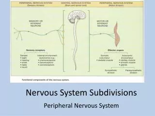
Nervous System Subdivisions. Peripheral Nervous System. Peripheral Nervous System. Cranial nerves arising from brain Somatic fibers connecting to skin & skeletal muscles Autonomic fibers connecting to viscera Spinal nerves arising from spinal cord

Nervous System Subdivisions
E N D
Presentation Transcript
Nervous System Subdivisions Peripheral Nervous System
Peripheral Nervous System • Cranial nerves arising from brain • Somatic fibers connecting to skin & skeletal muscles • Autonomic fibers connecting to viscera • Spinal nerves arising from spinal cord • Somatic fibers connecting to skin & skeletal muscle • Autonomic fibers connecting to viscera
Characteristics of Spinal Cord • 17” long; ¾” thick • 2-way conduction pathway ; to and from brain • Major reflex center • Extends from foramen magnum to L1 • Posterior to L1 is “cauda equina”
Gray Matter Looks like an “H” in cross section Contains: Posterior (dorsal) horn – axons of sensory neurons and association neurons Anterior (ventral) horn - cell bodies of somatic motor neurons
Amount of gray matter reflects amount of skeletal muscle innervated at that level Dorsal horn Ventral horn
Nerve Roots Dorsal nerve root – carries incoming messages (sensory) Ventral nerve root – carries outgoing messages (motor)
Dorsal and ventral roots join to form 31 “spinal nerves” Area of skin innervated by each spinal nerve = “dermatome” Muscle group innervated by each spinal nerve = “myotome”
White Matter Contain “nerve tracts” of myelinated and unmyelinated fibers; run in 3 directions: Up (ascending; sensory) Down (descending; motor) Side to side
Functions of Spinal Cord Conduit of nerve impulses to and from brain & brainstem Center for spinal reflexes
Reflex Arcs • Reflexes are automatic, involuntary responses to stimuli • Simple reflex arc • Sensory – motor • Most common reflex arc • Sensory – association - motor
Reflex Behavior Knee-jerk reflex Simple monosynaptic reflex Helps maintain upright posture
Reflex Behavior Withdrawal reflex Prevents/limits tissue damage
Reflex Arc Crossed Extensor reflex Crossing of sensory reflexes impulses within the reflex center to produce an opposite effect
Uses of Reflexes Used to determine neurological status/dmg Plantar reflex – flexes foot Babinski reflex – abnormal; dorsiflexion of great toe Biceps-jerk reflex – tap on inside of elbow causes flexing Plantar reflex Babinski reflex
Uses of Reflexes Triceps-jerk reflex – tapping short tendon of triceps close to insertion near tip of elbow; extends elbow Abdominal reflexes – stroking skin of abdomen causes contraction of abdominal muscles & umbilicus moves twd stimulated area
Uses of Reflexes Ankle-jerk reflex – tapping calcaneous tendon above insertion on calcaneous = plantar flexion
Tracts of Spinal Cord Ascending tracts conduct sensory impulses to brain Descending tracts conduct motor impulses to motor neurons reaching muscles & glands
Spinal Cord Injuries 1st few days of spinal injury: vertebrae are compressed/may break = action potentials in neurons which kills them Dying neurons rls calcium ions; activates tissue-degrading enzymes Inflammation Axons tear, myelin coatings are stripped off; vital connections bt nerves/muscles cut Tissue is unable to regenerate
Spinal Injuries Injuring nerve pathways = depressed cord reflex activity (spinal shock) – normal reflex activity may rtn Severed nerve fibers = permanent loss of fxn Blow, whiplash, rupture of vertebral disc = pain, weakness, muscular atrophy
Pathology Paralysis – loss of motor function Parathesias – loss of sensation Paraplegia – loss of lower limb function Quadraplegia – loss of all limb function
Hemiplegia – brain disorder resulting in loss of function on one side Flaccid paralysis – loss of muscle tone due to damage of ventral horn or root Spastic paralysis – damage to brain neurons; muscles are stimulated by spinal nerves
Poliomyelitis – “gray matter inflammation” – viral disease which destroys the ventral horn of the spinal cord; virus is contracted through contaminated water; Salk and Sabin vaccines prevent infection
Regeneration – stems cells in rodents: regain some ability to walk Spinal Injury - Treatments
Nerve & Nerve Fiber Classification • Sensory Nerves • Conduct impulses into brain or spinal cord
Nerve & Nerve Fiber Classification • Motor Nerves • Conduct impulses to muscles or glands
Nerve & Nerve Fiber Classification • Mixed (both sensory & motor) nerves • Contain both sensory nerve fibers & motor nerve fibers • Most nerves are mixed nerves • ALL spinal nerves are mixed nerves (except 1st pair)
Cranial Nerves O-O-O-T-T-A-F-V-G-V-A-H (structural)
Cranial Nerves • Designated: ‘CN’ • Given Roman numerals: I – XII • “Oh-Oh-Oh-To-Touch-And-Feel-Very-Good-Velvet-AH!” - structural • “Some say marry money but my brother says bad business marry money” – sensory, motor or both
Cranial Nerve I • Olfactory Nerve (CN I) • Sensory nerve • Fibers transmit impulses associated with smell
Cranial Nerve II • Optic Nerve (CN II) • Sensory nerve • Fibers transmit impulses associated with vision
Oculomotor nerve (CN III) Primarily motor nerve Motor impulses to muscles that: Raise eyelids Move the eyes Focus the lens Adjust light entering eye Some sensory Proprioceptors – chgs in muscles & tendons such as at joints Cranial Nerve III
Cranial Nerve IV • Trochlear Nerve (CN IV) • Primarily motor nerve • Motor impulses to muscles that move eyes • Some sensory • proprioceptors
Trigeminal nerve (CN V) Mixed nerve “3 Sisters”: (1) Opthalmic division Sensory from sfc of eyes, tear glands, scalp, forehead, & upper lids Cranial Nerve V
Cranial Nerve V • (2) Maxillary division • Sensory from upper teeth, upper gum, upper lip, palate, & skin of face • (3) Mandibular division • Sensory from scalp, skin of jaw, lower teeth, lower gum, & lower lip • Motor to muscles of mastication & muscles in lower floor of mouth
Cranial Nerve VI • Abducens nerve (CN VI) • Primarily motor nerve • Motor impulses to muscles that move eye • Some sensory • proprioceptors
Cranial Nerve VII • Facial Nerve (CN VII) • Mixed nerve • Sensory from taste receptors • Motor to muscles of facial expression, tear glands, and salivary glands
Cranial Nerve VIII • Vestibulocochlear nerve (CN VIII) • Aka acoustic & auditory nerve • Sensory nerve
Cranial Nerve VIII • 2 branches: • Vestibular branch • Sensory from equilibrium receptors of ear • Cochlear branch • Sensory from hearing receptors
Cranial Nerve IX • Glossopharyngeal nerve (CN IX) • Mixed nerve • Sensory from pharynx, tonsils, tongue, & carotid arteries • Motor to salivary glands & muscles of pharynx
Cranial Nerve X • Vagus nerve (CN X) • Mixed nerve • Somatic motor to muscles of speech & swallowing


