Download
1 / 8
80 likes | 123 Vues
Explore 3D atlases of normal and mutant embryo development, gene expression, and live imaging of craniofacial structures. Discover advanced imaging tools for accurate data analysis and visualizations in this cutting-edge research field.

E N D
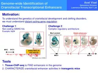
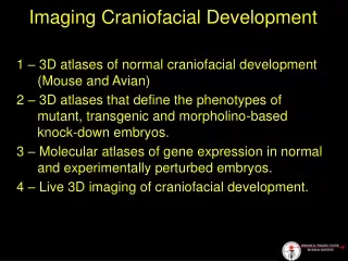
Imaging Craniofacial Development 1 – 3D atlases of normal craniofacial development (Mouse and Avian) 2 – 3D atlases that define the phenotypes of mutant, transgenic and morpholino-based knock-down embryos. 3 – Molecular atlases of gene expression in normal and experimentally perturbed embryos. 4 – Live 3D imaging of craniofacial development.
Volumetric Imaging Tools SIM OPT uMRI
Robust multiplexing of five target mRNAs in fixed whole-mount zebrafish H.M.T. Choi, J.Y. Chang, L.A. Trinh, J.E. Padilla, S.E. Fraser, N.A. Pierce, in review
Imaging Craniofacial Development 3-D imaging of anatomy volumetric data sets, tools 3-D imaging of gene expression volumetric data sets, tools 4-D imaging of development data sets, tools, pipeline Single cell in context is key