
Influence of Malignancy and Specimen Preparation on TRUS Biopsy Core Fragmentation
This study explores how the presence of malignancy and the method of specimen preparation affect TRUS biopsy core fragmentation. Results show differences in core length and number between benign and cancer patients. The discussion suggests a protective effect of swishing over swiping in cancer patients. References discuss the impact of core biopsy fragmentation on prostate cancer. Conclusions emphasize the importance of minimizing core fragmentation in cancer patients.

Influence of Malignancy and Specimen Preparation on TRUS Biopsy Core Fragmentation
E N D
Presentation Transcript
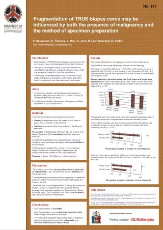
No. 117 Fragmentation of TRUS biopsy cores may be influenced by both the presence of malignancy and the method of specimen preparation P. Satasivam, R. Thomas, K. Rao, G. Jack, N. Lawrentschuk, D. Bolton The Austin Hospital, Heidelberg VIC • Introduction • Fragmentation of TRUS-biopsy cores is a phenomenon that has recently been acknowledged in the urologic literature. • The rate of core fragmentation is clinically relevant with regard to the identification of patients with low-volume CaP and subsequent selection for active surveillance. • Anecdotally, core fragmentation may be affected by the method of specimen preparation, with friction and subtle fractures occurring more often with certain techniques. • Results • There were 46patientsin the swipe group and 44 in the swish group. • 23 patients in each group had benign findings on final histology. • There was no significant difference in PSA (8.8 vs 9.0 ng/mL), mean core length (12.4 vs 13.4 mm) or final number of core fragments (15.0 vs 14.1) between the two groups. Each group had a similar number of patients with each Gleason score. • Cancer patients in theswish group had bothfewer and longer core fragments than those in the swipe group (See Table), a result which was repeated when analysed as a percentage increase in the number of core fragments, p = 0.03. • The patients within the swish group were then analysed separately, looking specifically at the rate of fragmentation within each sextant location. • The overall percentage core fragmentation among these 44 patients was significantly greater in benign sextant locations compared with those containing cancer, p = 0.017. • However, there was no significant difference in the fragmentation rate between benign and malignant locations in the 21 patients in the swish group with CaP. Aims To determine whether the technique used to prepare a prostate biopsy specimen affects the number or length of cores that are finally processed To determine whether the presence of malignancy affects the degree of core fragmentation Methods Two methods of specimen preparation compared: Swiping the specimen from the needle onto a piece of paper prior to transfer to the container Swishing the needle within the container to dislodge the specimen Prospective TRUS biopsies (minimum 12 core sextant) from May to December 2010 randomised to either swiping or swishing. All specimens processed at a single pathology laboratory using the same automated technique, pathologists blinded to randomisation. Pathology report analysed for number of cores reported, length of cores and histopathology of specimens and compared with the number of cores taken at biopsy. Powered to detect 15% difference between groups. Discussion TRUS biopsy cores from cancer patients have a lower rate of fragmentation , but only when the tissue is swished into the specimen jar. The act of swiping the biopsy specimen appears to negate this protective effect, most likely through friction that causes the cores to fracture. The similar rate of core fragmentation in benign and malignant sextant locations in patients with CaP suggests that the process that is protective against core fragmentation affects the entire gland. This “stickiness” may reflect scar formation after injury as in chronic inflammation. • References • Reis L, Reinato J, Silva D, Matheus W, Denardi F and Ferreira U: The impact of core biopsy fragmentation in prostate cancer. International Urology and Nephrology. 42: 965-969. • Klein EA and Silverman R: Inflammation, infection, and prostate cancer. Current Opinion in Urology. 18: 315-319 10.1097/MOU.0b013e3282f9b3b7, 2008. • Epstein JI, et al. “Nonpalpable stage T1c prostate cancer: Prediction of insignificant disease using free/total prostate specific antig en levels and needle biopsy findings.” J Urol, 1998: 160:2407-2411. • Conclusions • Core fragmentation is inevitable. • Core fragmentation may be minimisedin patients with CaPthrough modification of technique. • The finding that malignant tissue is less likely to fragment reinforces emerging theories of an inflammatory or infective aetiology for prostate cancer. Poster presentation sponsor


