CPC
CPC. David A. Farcy MD, FAAEM, FACEP, FCCM Chairman, Department of Emergency Medicine Mount Sinai Medical Center, Miami Beach , Florida. Case Summary. 59 y/o male with 4 days of congestion/fatigue/ exertional dyspnea/occasional palpitation. PMH: BPH, Vasovagal syncope

CPC
E N D
Presentation Transcript
CPC David A. Farcy MD, FAAEM, FACEP, FCCM Chairman, Department of Emergency Medicine Mount Sinai Medical Center, Miami Beach, Florida
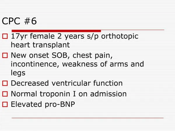
Case Summary • 59 y/o male with 4 days of congestion/fatigue/exertional dyspnea/occasional palpitation. • PMH: BPH, Vasovagal syncope • Medication: Rapaflo, OTC • Social: None contributory • Family: brother and cousin with pacemakers
Physical Exam: • Vital Signs – HR 42, BP 132/84, RR 14, O2 98% on RA, Temp 99.1F • AxOx3 • Rest of Exam none contributory.
ACLS • Need to decide if patient is stable or unstable. • In this case he relatively stable, without evidence of hypo perfusion. • WHAT is my EKG
EKG EKG Interpretation • 3rd degree Atrioventricular (AV)Block, with AtrioVentricular dissociation.
Other Results Lab X-ray and Echo • CXR: no acute finding, no cardiomegaly • CBC: 5.6> 16.4/49.6<195 • BMP: 144/3.4/103/34/15/1.0<92 • Troponin: <0.03 INR: 1.0
Course. • EP and Cardiology consulted with request for Echo and CTA heart • Echo: LVEF 55-60%, mildly dilated left atrium • CTA Heart: No coronary artery disease
Erroneous Dogma • A common misconception of an inexperienced clinician is to gauge a patient’s stability according to the heart rate and blood pressure rather than according to the symptoms and level of the block
Differential Diagnosis • Cardiac: ACS.. AVN block associated with inferior wall AMI, or His-Purkinje block associated with anterior wall AMI. • Medication interaction ( B-Blocker, Ca Channel blocker, Digoxin) • Infectious disease: Lyme disease, rheumatic fever, myocarditis, Chagas disease. • Metabolic causes - Hypoxia, hyperkalemia, hypothyroidism • Rheumatic diseases - Ankylosing spondylitis, Reiter syndrome, relapsing polychondritis, rheumatoid arthritis, scleroderma • Congenital: Lenegre’s – Lev disease,
Differential Diagnosis • Cardiac: ACS.. AVN block associated with inferior wall AMI, or His-Purkinje block associated with anterior wall AMI. Unlikely due to the fact: no ischemic changes on EKG, neg troponin, no calcification of CT.
Differential Diagnosis 2. Medication interaction ( B-Blocker, Ca Channel blocker, Digoxin) Unlikely, on none of those medication, only medication is rapaflo AV blocker not a know adverse reactions
Differential Diagnosis • Infectious disease: Lyme disease, rheumatic fever, myocarditis, Chagas disease. Unlikely: no report of fever, no reports of travel to any specific endemic area, no report of rash, joint pain
Differential Diagnosis 4. Metabolic causes - Hypoxia, hyperkalemia, hypothyroidism Unlikely: patient cxr normal, O2 sat normal, no electrolytes or renal disorder. Hypothyroidism could cause this but temp is normal and no history of temperature intolerance, no physical finding, so unlikely
Differential Diagnosis • Rheumatic diseases - Ankylosing spondylitis, Reiter syndrome, relapsing polychondritis, rheumatoid arthritis, scleroderma Unlikely. No joints involvement, no skin involvement, no murmurs, normal EF,
Differential Diagnosis • Congenital: Lenegre’s – Lev disease,
Lenegre's versus Lev's disease • Lenegre’s disease is idiopathic fibrosis of the cardiac conduction system resulting heart block. • Lev’s disease is calcification of the cardiac conduction system resulting in heart block. • This most commonly occurs as a part of the aging process of the heart in the elderly, however familial forms have been identified. • Stokes-Adams attacks can be associated with Lenegre’s disease and result in syncope. • Treatment is a permanent pacemaker depending on the rhythm that manifests.
DIAGNOSIS • LENEGRE-LEV Disease because: • Hx pacemaker in brother and cousin • Older population patient is 59 y.o • Hx of vasovagal syncope. Need permanent pacemaker