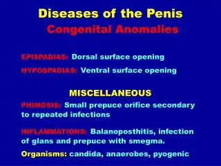
In general the origin of the problems or diseases in the motion system can be - Congenital - Tumor
In general the origin of the problems or diseases in the motion system can be - Congenital - Tumor - Inflammation - Trauma - Arthrosis (osteoarthritis) = degenerative joint disease. HEAD Configurations of the cranium: - narrow in the frontal plane - wide in the frontal plane

In general the origin of the problems or diseases in the motion system can be - Congenital - Tumor
E N D
Presentation Transcript
In general the origin of the problems or diseases in the motion system can be -Congenital -Tumor -Inflammation -Trauma -Arthrosis (osteoarthritis) = degenerative joint disease
HEAD Configurations of the cranium: -narrow in the frontal plane -wide in the frontal plane -cephalhaematoma external -disturbances of ossification, craniosynostosis
The ossification of the cranial bones takes place in a desmogen way; the collagen tissue colony change directly into bone. At the beginning the bone is growing from the ossification center in radial direction and takes a star-like form with toothed edges. The toothed edges come together until the teeth fit into each other. Further on, the bony growth is continued with primer angiogen ossification.
The craniosynostosis is an irregularity of development resulting from the early (premature) ossification of the immovable joints between the cranial bones.
The position of the head and its motion will determine basically the development of the posture of human body. One of the main characteristics of human posture and motion is the flexible and balanced control of the head on the top of the vertically positioned spine. The development of head control is also in connection with the development of the three dimensional position of the trunk as well as the stability of spine.
In case of a matured new-born in supine position, the contraction of occipital/cervical muscle moves the head upwards for 1 –to 2 seconds; still, it is not a spontaneous raising of the head. Normally, in supine position, the raising of head can be observed from the third week, that is usually associated with movements of the trunk and extremities.
Hypotonic? In case of functional insufficiency and reduced tone of axial muscles, the head remains leant back and the trunk is bended backwards in lordosis.
THORACIC DEFORMITIES Etiologic: genetical determination ( mesenchyme germ colony) Dominant or recessive type of inheritance The excessive growth of the ribs is the dominant factor in the deformity.
Accompanying deformities: Marphansy., heart-vitium, scoliosis ( The latter shall be distinguished from the consequential deformity of the chest in scoliosis! )
Itsformsare: A. Depresseddeformities Pectusexcavatum - centralsymmetric - asymmetric - centralsymmetric + flat back – syndrome - asymmetric + flat back - syndrome B. Protrusiontype - Pectuscarinatum (manubrio-gladiolar-type) - Pectusarcuatum (chondro-manubrial - type) C. Pectusdeformatum ( longitudinal, one-ordoublesided)
In the first decade the inflammation of the breathing tubes, the paradox breathing is the most common problem. Between 10 and 20 dyspnea, palpitation, ES occurs, and psychic problems increasing with the age.
Gymnastics. swimming? • Respiration: • articular and osseousmechanisms • respiratorymuscles (mm.intercostales, diaphragm) • respiratoryassistingmuscles
The area of the chest dilates or decreases as the ribs turning around their joint axis. This is the articular and bony mechanism of breathing.
The sternal part of diaphragm is weak, the pars costalis arises from the cartilaginous surface of the last six ribs and is directed upwards vertically and then inwards.
Aspiratoryassistingmuscles: with fixed armsthethoracohumeralmusclesarisingfromtheribs. M.pectoralis maior et minor, M. serratusanterior, M. latissimusdorsi. Mm. Scaleni, M.sternocleidomastoideus
Conservative treatment can be required: • if regression is expected • before surgery • Postoperative treatment, physiotherapy to improve the posture • When surgery is contraindicated
Shoulders falling forwards, accompanying deformity of spine, paradox movement of the frontal wall of the thorax lead to the conclusion that progression exists. Physiotherapy: respiratory exercise muscle strengthening exercise to improve the posture
The respiratory exercise is successful if it shows and exercises how to use the thoracic and abdominal components of breathing separately in a conscious way. Swimming – provides assistance in attaining the proper and economic respiratory technique.
Indications for surgical treatment: -severe deformity -pectus-index below 30% -cardiac problems -impairment of respiratory function
„Pectus- index”: the smallest internal sternovertebral distance (measured on the punctum maximum of the impression) divided by the thoracic transversal diameter (distance between the inner sides of the right- and left walls of the chest (thoracic cavity) measured at the height of diaphragm x 100. The index above 35% is normal, between 30 and 35% means mild decreasing of the space. In the cases of protrusion type deformities the index is between 45 and 60% .
The hip—joint is the most proximal element of the kinetic chain of the lower extremity.In mechanical respect, the hip is the simplest joint – a ball- and-socket joint, where the joint head is the femur head while the socket is the acetabulum.The input plane the acetabulum forms an angle between 30 and 35 degrees with the sagittal plane and an angle between 45 and 50 degrees with the horizontal plane.While the circular fibrous cartilage (labrum) on the edge of the acetabulum encloses the femoral head beyond its equator the hip joint is really an enarthros.
In standing position, the body weight is distributed equally on the two hip joints. Under normal anatomic conditions, the line of action of pressure force passes through the centre of head and is perpendicular to the surface of them. In the stance phase during gait, however, it is only the supporting lower extremity that bears the weight of body and trunk, resp.Pauwels wrote that the weight force of the body acts medially from the hip joint. The lever of body weight is approx. 3-times longer than that of the load that is the lever of muscle.
Consequently, to maintain the horizontal equilibrium of the pelvis a muscle force about three times as much as the body weight is required. The resulting force acting on the femur head is given by the vector sum of the acting forces, that results in a force four times as much as the body weight.
During the intrauterine growth, the hip joint developed in the 10th week old embryo.
The form of the acetabulum is varying from the intrauterine life onwards up to the age 15 – 18.On the first two-third part of fetal life, it is dome-shaped and, then, its depth is gradually decreases and, at the time of birth, the vertical diameter is only two-fifth of the horizontal diameter. After birth, it is further deepened. The completely developed acetabulum takes the shape of a spherical segment of 170 to 175º.
During the development of hip joint, the collo-diaphyseal angle and the antetorsion of the femoral neck are varying The collo-diaphyseal angle is the angle formed by the femoral neck with the longitudinal axis of the femoral shaft in the frontal plane. The anteversion/antetorsion is the angle formed by the femoral neck with the femur condyles in the frontal plane.