Download
1 / 70
760 likes | 934 Vues
Incredible Nervous System. http://thebrain.mcgill.ca/flash/index_i.html. STUDYING THE LIVING BRAIN. Brain scans techniques that can look through the thick skull and picture the brain with astonishingly clarity yet cause no damage to the extremely delicate brain cells.

E N D
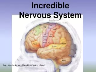
Incredible Nervous System http://thebrain.mcgill.ca/flash/index_i.html
STUDYING THE LIVING BRAIN • Brain scans • techniques that can look through the thick skull and picture the brain with astonishingly clarity yet cause no damage to the extremely delicate brain cells
STUDYING THE LIVING BRAIN • EEG: • Electroencephalogram: studies the different electrical brain waves generated by neurons. Gives a computerized read-out showing the activity on the brain’s surface.
CT/CAT Scans http://www.youtube.com/watch?NR=1&v=rN4E8Y5loAs
STUDYING THE LIVING BRAIN • MRI • magnetic resonance imagery • involves passing nonharmful radio frequencies through the brain • fMRI • functional magnetic resonance imaging • measures the activity of specific neurons that are functioning during cognitive tasks, such as thinking, listening
http://www.youtube.com/watch?v=9E1GoWhSlho&feature=related MRI http://www.youtube.com/watch?v=Cwda7YWK0WQ fMRI http://www.youtube.com/watch?v=BmQR57V5TVU&feature=related http://www.youtube.com/watch?v=uVht8AMknfc&feature=related http://www.youtube.com/watch?v=AUT9UTVrwp8&feature=related
STUDYING THE LIVING BRAIN 3.PET scans • positron emission tomography • involves injecting a slightly radioactive solution into the blood and then measuring the amount of radiation absorbed by neurons • http://www.youtube.com/watch?NR=1&v=d9iOxMFmPlA
transcranial magnetic stimulation http://www.sciencedaily.com/releases/2005/10/051019003056.htm
ORGANIZATION OF THE BRAIN …we will look at • Major divisions of the nervous system • central nervous system - CNS • peripheral nervous system - PNS
PERIPHERAL & CENTRAL NERVOUS SYSTEM …remember • Peripheral Nervous System • made up of nerves that are located throughout the body, except in the brain & spinal cord • Central Nervous System • made up of neurons located in the brain & spinal cord
ORGANIZATION OF THE BRAIN • Peripheral nervous system - PNS • includes all the nerves that extend from the spinal cord and carry messages to and from various muscles, glands, and sense organs located throughout the body • Subdivisions of the PNS • somatic nervous system • autonomic nervous system - ANS • sympathetic division • parasympathetic division
ORGANIZATION OF THE BRAIN • Somatic nervous system • network of nerves that connect either to sensory receptors or to muscles that you can move voluntarily, such as muscles in your limbs, back, neck, and chest • nerves contain two kinds of fibers • Afferent • sensory fibers; carry information to the brain • Efferent • motor fibers; carry information from brain or spinal cord to the muscles
ORGANIZATION OF THE BRAIN • Autonomic nervous system - ANS • regulates heart rate, breathing, blood pressure, digestion, hormone secretion, and other functions A. Sympathetic division • triggered by threatening or challenging physical or psychological stimuli, increases physiological arousal and prepares the body for action (fight/flight) B. Parasympathetic division • returns the body to a calmer, decreases physiological arousal(decreases heart rate/lowers BP) and is involved in digestion • Homeostasis: • sympathetic and parasympathetic systems work together to keep the body’s level of arousal in balance for optimum functioning
ORGANIZATION OF THE BRAIN • Major Parts of the Brain • Forebrain 2.Midbrain 3.Hindbrain
ORGANIZATION OF THE BRAIN • Forebrain • largest part of the brain • has right and left sides called hemispheres • hemispheres are responsible for a number of functions, including learning and memory, speaking and language, emotional responses, experiencing sensations, initiating voluntary movements, planning, and making decisions
The Cerebral Cortex • Four lobes • Frontal lobe • Parietal lobe • Occipital lobe • Temporal lobe Each hemisphere of the cerebral cortex is divided into four lobes
Wrinkled cortex a thin layer of cells that essentially covers the entire surface of the forebrain the evolutionary purpose of the outer cortex being wrinkled is that it is able to contain billions of more neurons than if it was a smooth surface CONTROL CENTERS: FOUR LOBES
Frontal Lobe overview: involved with personality, emotions, and motor behaviors • THE CEREBRUM:Frontal Lobe • Behavior • Abstract thought processes • Speech • Attention • Intellect • Reflection • Initiative • Coordination of movements • Muscle movements • Skilled movements
Frontal lobe: functions motor cortex narrow strip of cortex that is located on the back edge of the frontal lobe and extends down its side involved in the initiation of all voluntary movements right side controls left left side controls right organization and function of motor cortex Frontal Lobe
Language in the Brain http://www.news-medical.net/health/The-Human-Brain.aspx
Parietal Lobe Overview: involved with perception and sensory experiences • location of somatosensory cortex:narrow strip of cortex that is located on the front edge of the parietal lobe and extends down its side • Sense of touch (tactile sensation) • Appreciation of form through touch • Response to internal stimuli • Sensory combination and comprehension • Some language and reading functions • Spatial associations
Parietal Lobe • Parietal lobe: function • involved in several cognitive functions, including recognizing objects, remembering items, and perceiving and analyzing objects in space
Temporal Lobe Overview:involved with hearing and speaking • Temporal Lobe • Auditory memories • Some hearing • Music • Fear • Language • Speech • Emotions
Temporal lobe: functions primary auditory cortex located on top edge of each temporal lobe, receives electrical signals from receptors in the ears and transforms these signals into meaningful sound sensations, such as vowels and consonants auditory association area:located directly below the primary auditory cortex transforms basic sensory information, such as noises or sounds, into recognizable auditory information, such as words or music Temporal Lobe
Temporal Lobe • Temporal lobe: functions • Broca’s area - frontal lobe • located in left frontal lobe • necessary for combining sounds into words and arranging words into meaningful sentences • Wernicke’s area • located in the left temporal lobe • necessary for speaking in coherent sentences and for understanding speech
Occipital Lobe • Overview: involved with visual processing • Vision • Reading • Visual memory and associations
Occipital Lobe • Occipital lobe: functions • Vision: primary visual cortex which is: located at the very back of the occipital lobe • receives electrical signals from receptors in the eyes and transforms these signals into meaningless basic visual sensations, such as lights, lines, shadows, colors, and textures • visual association area: • transforms basic sensations, such as lights, lines, colors, and textures, into complete, meaningful visual perceptions, such as persons, objects, or animals
Functional Areas of the Brain Lobe Review Slide Functional Areas of the Brain http://www.umich.edu/~cogneuro/jpg/Brod_sagtl.gif
Forebrain: Limbic System: The Old Brain The Limbic System though very basic and old constitutes a part of the Forebrain. • Structures: • Hypothalamus • Amygdala • Thalamus • Hippocampus
Hypothalamus regulates many motivational behaviors, including eating, drinking, and sexual responses; emotional behaviors such as arousing the body when fighting or fleeing, and secretion of hormones, such as occurs at puberty Amygdala located in the tip of the hippocampus receives input from all the senses evaluates the emotional significance of stimuli and facial expressions, especially those involving fear, distress, or threat Forebrain: Limbic System: Old Brain
3. Thalamus gathers and processes information from the senses involved in receiving sensory information, doing some initial processing, and then relaying the sensory information to areas of the cortex 4. Hippocampus curved structure inside the temporal lobe Involved in saving many kinds of fleeting memories by putting them into permanent storage in various parts of the brain LIMBIC SYSTEM: OLD BRAIN
Midbrain/Mesencephalon has areas for vision, hearing, eye and body movement contains the reticular formation, which arouses the forebrain so that it is ready to process information from the senses essential for processing voluntary motor movement 2. VTA: mechanism greatly involved in the feeling of pleasure 3. Nucleus Accumbens: same as VTA (these neurons are linked with the VTA) Midbrain