Download
1 / 63
640 likes | 839 Vues
CATABOLISM OF PROTEINS AND AMINO ACIDS. Prof.Dr.Arzu SEVEN. In animals,amino acids undergo oxidative degradation in 3 different metabolic circumstances:

E N D
CATABOLISM OF PROTEINS AND AMINO ACIDS Prof.Dr.Arzu SEVEN
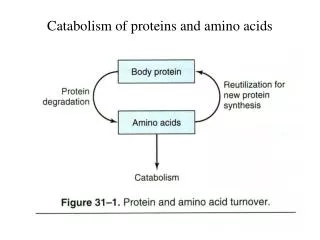
In animals,amino acids undergo oxidative degradation in 3 different metabolic circumstances: • 1-During normal synthesis and degradation of cellular proteins, some amino acids, that are not needed for new protein synthesis, undergo OXİDATİVE DEGRADATİON • 2-When a diet is rich in protein, the surplus amino acids are catabolized ( in the liver amino acids can't be stored) • 3-During starvation and uncontrolled DM, when carbohydrates are unavailable or improperly utilized, cellular proteins are used as fuel.
Animals convert α-amino nitrogen to varied end products as ammonia, uric acid or urea. • Humans are ureotelic and excrete non-toxic, water-soluble urea.
BİOSYNTHESİS OF UREA • Urea biosynthesis occurs in 4 states • 1-Transamination • 2-Oxidative deamination of glutamate. • 3-Ammonia transport • 4-Reactions of urea cycle.
Transamination • Transamination transfers α-amino nitrogen to α-ketoglutarate, forming glutamate. • Transamination interconverts pairs of α-amino acids and α-ketoacids.
Amino acids that don't participate in transamination: • Lysine, threonine, proline, hydroxyproline. • Reversible • Aminotransferases (transferases) remove the amino group from most amino acids and produce the corresponding α -ketoacid
Cofactor:Pyridoxal phosphate • Pyridoxamine is the intermediate in the reaction. • Alanine-pyruvate amino transferase (alanine aminotransferase) and glutamate α-ketoglutarate amino transferase (glutamate aminotransferase) catalyze the transfer of amino groups to pyruvate (forming alanine) or to α-ketoglutare (forming glutamate)
Each aminotransferase is specific for one pair of substrates but nonspecific for the other. • Since alanine is also a substrate for glutamate aminotransferase, all the amino nitrogen from amino acids that undergo transamination can be concentrated in glutamate
The effect of transamination reaction is to collect the amino groups from many different amino acids in the form of L-glutamate. • L-glutamate then functions as an amino group donor for biosynthetic pathways or for excretion pathways that lead to the eliminaton of nitrogenous waste products.
Glutamate releases its amino qroup as ammonia in the liver. • In hepatocytes, glutamate is transported from cytosol into mitochondria, where it undergoes OXİDATİVE DEAMİNATİON by glutamate dehydrogenase.
L-glutamate is the only amino acid that undergoes oxidative deamination at an appreciable rate in mammalian tissues. • L-glutamate dehydrogenase (GDH) occupies a central position in nitrogen metabolism.(mitochondrial matrix)
GDH reaction is a reversible reaction that can produce glutamate from α-ketoglutarate or convert glutamate to α-ketoglutarate and NH3 • Hepatic GDH can use either NAD+ or NADP+, as the acceptor of reducing equivalents. • Glutamate serves as a precursor of ammonia.Mitochondrial glutamine synthetase catlyses this energy requiring reaction (ATP), consuming a molecule of ammonia.
Glutamine synthetase is a primary regulatory point in nitrogen metabolism. • It is regulated both allosterically and by covalent modification (adenylation inactivation).
Glutamine can serve as a buffer for ammonia utilization as a source of ammonia and carrier of amino groups. Glutamine, along with alanine, is a key transporter of amino groups between various tissues and liver and is present in greater concentrations than most amino acids in blood.
Glutaminase hydrolyses glutamine to glutamate and NH4+. • This reaction is important in the kidney for the management of proton transport and pH control.
Aminotransferase + GDH action TRANSDEAMiNATiON • GDH operates at an important intersection of carbon and nitrogen metabolism. • The mammalian GDH is allosterically regulated by GTP (-modulator) and ADP (+ modulator)
Hyperinsulinism-hyperammonemiasyndrome: • Mutations that alter the allosteric binding site for GTP • Permanent activation of GDH • Genetic disorder • NH3 increase (in blood) • Hypoglycemia
Oxidative deamination of amino acids • L-amino acid oxidases of liver and kidney produces NH3 and α-keto acid directly, using FMN as a cofactor.(through α-imino acid) • FMNH2 is converted to FMN, using O2 and produces H2O2 which is decomposed by catalase.
Non-oxidative deamination • Hydroxyaminoacids (serine, threonine) are non-oxidatively deaminated by dehydratase to form keto acids (pyruvate, and α-ketobutyrate) and NH3.
The NH4+ ,from intestine and kidney, is transported in the blood to liver. • In the liver, the ammonia from all sources is disposed of by urea synthesis.
Ammonia Transport • Ammonia produced by enteric bacteria and absorbed into portal venous blood and ammonia produced by tissues are rapidly removed from circulation by liver and converted to urea. • Only traces (10-20 μg/dl) are normally present in peripheral blood.
This is essential since NH3 is toxic to central nervous system. • In severely impaired hepatic function and in the development of collateral links between portal and systemic veins ,cirrhois,ammonia intoxication develops.
Symptoms:Tremor, slurred speech, blurred vision, coma • Ammonia Encephalopathy • When ammonia concentration increases in blood and other biological fluİds, it diffuses across blood-brain barrier.
Increased synthesis of glutamate from α-ketoglutarate leads to α-ketoglutarate depletion in CNS cells, resulting in TCA cycle inhibition and ATP decrease. • Glutamate, a major inhibitory neurotransmitter, or its derivative GABA, may also contribute to CNS effects.
The sensitivity of brain to ammonia may reflect the depletion of neurotransmitters as well as changes in cellular osmotic balance. • GDH and glutamine synthetase are present at high levels in the brain, although glutamine synthetase reaction is the more important pathway for removal of NH4+. • High levels of NH4+ lead to increased levels of
glutamine, which acts as an osmotically active solute (osmolyte) in brain astrocytes. • Uptake of water into astroyctes to maintain osmotic balance leads to swelling of cells and coma. coma
NH3 may be toxic to brain because it reacts with α-ketoglutarate to form glutamate. • Depleted levels of α-ketoglutarate impair TCA cycle function. • Excretion into urine of ammonia produced by renal tubular cells facilitates cation conservation and regulation of acid-base balance. • NH3 production from intracellular renal glutamine increases in metabolic acidosis, decreases in metabolic alkalosis.
Urea Cycle • Urea is the principal nitrogenous excretion product in humans. • The urea cycle was the first metabolic cycle to be well defined. • Its description preceded that of TCA cycle.
Synthesis of 1 mol. of urea requires 3 mol. of ATP (4 high energy phosphate groups) plus 1 mol. of ammonium and of α-amino nitrogen of aspartate. (Source of nitrogen atom) • Of the 6 participating amino acids, N-acetylglutamate functions only as an enzyme activator.
Ornithine, consumed in reaction 2, is regenerated in reaction 5. • There is no net loss or gain of ornithine, citrulline, argininosuccinate or arginine. • Ammonium ion, CO2 , ATP and aspartate are consumed. • Some reactions occur in the matrix of mitochondrion and others in the cytosol of the liver.
The start of urea cycle is the synthesis of carbamoyl phosphate from an ammonium, derived primarily from glutamate via GDH, and CO2 ( as bicarbonate) produced by mitochondrial respiration in liver. • This reaction requires 2 molecules of ATP and is catalyzed by carbamoyl phosphate synthetase I (CPS I) ,rate limiting enzyme of the urea cycle.
.CPS 1 requires N-acetylglutamate as a cofactor. • CPS 2, found in the cytosol, is involved in pyrimidine biosynthesis and does not require N-acetylglutamate, uses glutamine rather than ammonia as the nitrogen donor • 1 mol of ATP serves as a phosphate donor
Conversion of the second ATP to AMP and pyrophosphate, with the hydrolysis of pyrophosphate to ortophosphate, • provides the driving force for the synthesis of the amide bond and the mixed acid anhyride bond of carbamayl phosphate. • (high group transfer potential)
Ornithine transcarbamoylase catalyses the condensation of carbamoyl phosphate with amino acid ornithine to form citrulline. • Ornıthine plays a role resembling that of oxaloacetate in citric acid cycle, accepting material at each turn of cycle.
Citrulline passes from mitochondrion to cyctosol and condenses with • aspartate to form argininosuccinate. • This step is catalyzed by argininosuccinatesynthetase and requires ATP. • The reaction cleaves ATP to AMP and PP • which is hydrolyzed to two phosphate.
The formation of argininosuccinate provides the second nitrogen of urea. • Argininusuccinate is cleaved by argininosuccinase(reversible) to arginine and fumarate.
Cleavage of arginine by arginase (cytosolic) releases urea and reforms ornithine.
Ornithine reenters liver mitochondria for new urea synthesis. • Ornithine and lysine are potent inhibitors of arginase. • Urea diffuses into the blood, is transported to kidney and excreted in urine.