Download
1 / 52
550 likes | 948 Vues
The cellular basis of reproduction and inheritance. Chapter 8. How do cells divide? Is this important?. Organisms are made of cells. All cells come from cells. Cells reproduce. Mitosis makes new cells. This is asexual reproduction. Organisms reproduce. Meiosis is sexual reproduction.

E N D
The cellular basis of reproduction and inheritance. Chapter 8
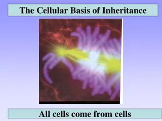
How do cells divide? Is this important? • Organisms are made of cells. • All cells come from cells. • Cells reproduce. • Mitosis makes new cells. • This is asexual reproduction. • Organisms reproduce. • Meiosis is sexual reproduction. • Meiosis ensures diversity. • Some cells are programmed to die. • Apoptosis destroys cells. • This keeps you from growing forever. This is an actual cell that has split its DNA, and is ready to split in half.
In a general sense, “like begets like.” • Asexual reproduction • When a cell makes exact copies of itself. • This provides “genetic uniformity.” • When you cut your skin, you want new skin cells to form, which are all alike. • Sexual reproduction • When a new cell is formed from the combination of two different cells. • This combination between two genetically distinct cells produces offspring that are also unique.
Cells arise only from pre-existing cells. • This is one of the main features of the “cell theory.” • Cell division is the heart of reproduction. • Cells don’t magically arise or appear. • They need to inherit genetic material. • But cells would take over the earth, if they kept reproducing. • Cell death (apoptosis) balances cell production.
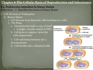
Prokaryotes reproduce by “binary fission.” • Features of prokaryotes? • DNA is small, and circular. • They usually produce asexually. • Binary fission is “simple” division: • The cell makes a copy of its DNA. • DNA is pushed to either end of cell. • It then “pinches off” forming two new cells. • This is somewhat basic, and you end up with two identical daughter cells. • Why is this called asexual reproduction?
Is cell division in your cells this easy? • Think about differences between eukaryotes and prokaryotes. • Eukaryotes have more complicated DNA. • Eukaryotic DNA is found in chromosomes. • The chromosomes contain the DNA, and must make a copy of themselves, in order to put DNA into each new cell.
What is a chromosome? • Chromosomes are molecules that contain DNA. • DNA contains your genes which is what you are. • How many chromosomes do you have? • 46 (or 23 pairs). • Each pair receives one from your mother, and one from your father. • When the chromosome makes a copy of itself, it has two halves, called chromatids. • These are connected at the centromere.
DNA is where your genes are. Your chromosomes “hold onto” the DNA. • In essence, the DNA is what is important, but the chromosome is where you find the DNA. • All of your genes are located on the 46 chromosomes.
When chromosomes do make a copy of themselves, the two “parts” are connected at a centromere.
How does a cell know when to make a copy of its DNA? • A cell will not make new DNA unless it is preparing to split into two daughter cells. • In a cells “life” there are times when the cell is just doing its work, and there may be a time when the cell “needs” to divide in half.
The cell cycle is divided into “parts” depending on what the cell is doing. • Interphase • Makes up 90% of the life cycle. • The cell is doing its work, or making a copy of its DNA. • Mitosis • Actual cell division. • Steps needed to divide the chromosomes in half. • Eukaryotes have a nucleus and chromosomes, and the cell needs to deal with these.
Interphase is a time of great activity. • A cell in interphase is “living its life.” • If the cell is a liver cell, then it is acting like a liver cell. • It could be growing, maturing, or doing its work. • This usually makes up 90% of the cells life cycle. • When the cell “decides” it needs to divide, only then does it go into mitosis. • Interphase is divided into three main phases: • “G1” phase. • A new cell after being formed by mitosis. • The cell is growing, maturing, and performing its work. • “S” phase. • This is called the “synthesis” phase. • The cell has decided that it needs to divide, therefore it needs to synthesize new DNA. • All of the cells energy is spent making a copy of its DNA, forming chromatids. • “G2” phase. • This is the cell, after DNA has been replicated. • The cell makes final preparations for cell division (mitosis).
Interphase • When you look at a cell in interphase, you will see: • The nucleus • The nucleolus inside of the nucleus. • DNA which looks “grainy” inside of the nucleus. • What you won’t see: • The DNA dividing. Plant cell (above) and animal cell in interphase.
Once the cell has replicated its DNA, it now needs to actually divide through mitosis. • The cell must first divide the chromatids on the chromosome. • After the chromosomes are split apart, then the cell can divide as well.
What happens during prophase? • The chromosomes, have become visible. • They were loose chromatin, and have twisted up to become thicker. • The centrioles have separated. • Spindle fibers grow, forcing the centrioles to move to opposite “poles” of the cell. • These fibers will eventually pull the chromosomes apart. • The nuclear membrane has broken apart. • This allows the spindle fibers to attach to each of the chromosomes.
What happens during metaphase? • The spindle fibers align the chromosomes along the “metaphase plate.” • If the centrioles are at the “poles” then the metaphase plate is at the “equator.” • It does this to prepare to pull the chromatids apart, to separate chromosomes. • This is usually easy to see in the microscope.
Metaphase in plant and animal cells. Notice the “plate” has chromatids hanging above and below it, but you can still see the metaphase plate. This is a very well-defined plate.
What happens during anaphase? • The centrioles pull the spindle fibers. • This pulls apart the chromatids. • The centromere region is split. • As soon as the chromatids are separate, they are considered distinct chromosomes. • The chromosomes are now pulled towards opposite ends of the cell. • Notice how they look like a “V” shape. • Why do you think they look like this?
Plant and animal cells in anaphase. Sometimes you will see the chromosomes closer to each other, sometimes they are further apart (like in these photographs).
What happens during telophase? • The chromosomes are now at opposite ends of the cell. • The cell membrane pinches inward in animal cells. • This is called a “cleavage furrow.” • In plant cells, a “cell plate” forms. • The cell is preparing to divide itself.
Telophase in plant and animal cells. Notice the cell plate forming in the center of the cell. This will form the new cell wall. Without this cell plate, the cell still is in anaphase. Notice the membrane forming a cleavage furrow. The red arrow indicates the new membrane between daughter cells. Notice how they are “pinched” above and below the arrow.
What is cytokinesis? • This is when the cytoplasm is actually dividing. • This is at the end of telophase. • At the end of cytokinesis, two new daughter cells are formed. • Each are now at G1 of interphase.
How can you learn these? • You must be able to identify the various stages of the cell cycle for lab. • Both plant and animal cells. • Make sure that you know what happens during each step of interphase and mitosis. • This takes patience, and practice. • Only using the microscope will help. • The cells don’t always look like the photographs.
That is how cells divide! • You have started with one cell: • End up with two identical daughter cells. • Each has an exact copy of the parent DNA. • This is why all cells come from pre-existing cells, and is part of the “cell theory.” • This process wasn’t understood until recently.
Why don’t cells keep dividing forever? • If they did, what would happen? • If a 8μm3 bacterial cell divided every twenty minutes: • In 36 hours it would cover the Earth 1 ft deep. • In 36 hours, 20 minutes? Two feet deep. • What factors limit cell growth? • Does any cell keep dividing like this?
Things that stop cell growth: • Actual control of the cell cycle. • Destruction of telomeres. • Contact inhibition. • Also called “density dependent inhibition.” • Chemical or molecular regulation. • Actual cell death. • Programmed or accidental.
The control of the cell cycle: • There are “checkpoints” along the way, during the cell cycle. • If something is wrong at any checkpoint, the cell won’t divide.
Role of telomeres in cell reproduction: • Telomeres are located at the end of the chromosome. • They form an enzyme called telomerase. • Basically speaking, if the telomeres wear out, the cells won’t divide.
Contact Inhibition • One additional thing that keeps cells from dividing is when they get crowded, they stop reproducing. • This is called contact inhibition. • Imagine cells that don’t stop dividing. • The would continue to split. • They would form a “mass” of cells. • This is how cancer begins.
Programmed cell death: • Its hard to imagine that certain cells are programmed to die. • This is part of normal embryological development. • Apoptosis • Rapidly and neatly dismantles cells. • Necrosis • Very messy! • Cell swells and bursts. • Inflammation • “Necrotic tissue”
What about cancer? • Cancer cells continue to divide. • They don’t stop. • Contact Inhibition? • They may take many years to spread. • Evidence shows that cancer cells actually attract blood and nutrients.
Causes of cancer:Mitosis in the absence of apoptosis. • Oncogenes • Genes that promote division which are overproduced. • New cells can form a tumor. • Tumor suppressor genes • Genes which prevent cell growth. • Inactivation of this “suppressor” gene causes cancer. • Environmental conditions • Diet, exercise, sun exposure, smoking, chemicals, radiation.
We have completed mitosis. Now lets concentrate on sexual reproducion. • Mitosis occurs in somatic cells. • Makes two daughter cells that are identical. • Meiosis occurs in sex cells. • Makes up to four cells that are genetically distinct.
Meiosis is “sexual reproduction” because it mixes up genes. • Genes can be mixed in two different ways: • Two different parents contribution their genes to their offspring. • An individual “shuffling” their genes, like a deck of cards. • Both of these contribute to “genetic diversity” which is essential. • Are you just like your parents? • Why or why not?
Some terms in meiosis you need to know. • Sex cells • The cells in your gonads that make sperm or eggs. • Somatic cells • All of the cells in your body, except for your sex cells. • Sex chromosomes • The “X” or “Y” chromosomes that determine your sex. • “XX” if your are female, or “XY” if you are a male. • Autosomes • The remaining 44 chromosomes that both males and females have. • Gametes • The sex cells (sperm or eggs) that contribute to an offspring. • Zygote • The fertilized egg which has both sets of chromosomes. • Diploid • A cell with the full compliment of chromosomes (46 in your cells). • This is referred to as 2n. • Haploid • A cell with half of its normal number of chromosomes. • Sperm and eggs each have 23 chromosomes. • This is referred to as 1N.
After replication, chromosomes are matched as “homologous” pairs. • Remember, you have 23 pairs of chromosomes. • These homologous chromosomes align into “tetrads.” • There are two chromosomes. • Each homologous pair (tetrad) has four chromatids and two chromosomes. There are two tetrads here.
Meiosis reduces the number of chromosomes in half. • From diploid to haploid. • 2N to 1N. • In you, from 46 to 23. • When a zygote is formed, it restores the 2N condition. • That way your baby has 46 chromosomes. • You will not have to identify meiosis under a microscope, but you need to understand how it is different than mitosis.
Prophase in mitosis Chromosomes become visible. Centrioles move to opposite poles. Prophase I in meiosis Chromosomes become visible. Centrioles move to opposite poles. Homologous chromosomes form tetrads. Crossing over occurs. Prophase I is the major difference between mitosis and meiosis.
Homologous chromosomes can carry different gene versions. • Homozygous chromosomes • These carry the same “kind” of genes. • Heterozygous chromosomes • These carry “different kinds” of genes.
Tetrads form during Prophase I. • Tetrad formation is important. • Tetrads allow the cell to go from 2N to 1N. • Tetrads allow crossing over to occur. • This increases genetic diversity greatly. • No two eggs or sperm are the same. • No two offspring are the same.
Crossing over adds additional genetic variation. • Imagine, DNA actually being “swapped” between chromosomes. • This occurs in Prophase I of meiosis. • Genetic diversity is the key to meiosis, and this diversity is amazing.
Metaphase I aligns the tetrads along the metaphase plate.This is strictly by chance… • This is called independent orientation or independent assortment. • Notice the two scenarios: • On the left, the tetrads lead to blue or green gametes. • On the right, which occurs half of the time, you have different gametes. • How do you think this leads to genetic diversity?
A karyotype is a “picture” of your chromosomes. • These are useful tools to determine normal and abnormal chromosome numbers. • What kinds of conditions can be diagnosed with a karyotype? • What is an amniocentesis?