Download
1 / 31
340 likes | 705 Vues
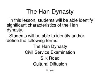
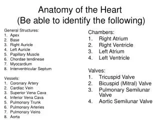
Anatomy of the Heart (Be able to identify the following). General Structures: Apex Base Right Auricle Left Auricle Papillary Muscle Chordae tendineae Myocardium Interventricular Septum Vessels: Coronary Artery Cardiac Vein Superior Vena Cava Inferior Vena Cava Pulmonary Trunk

E N D
Anatomy of the Heart (Be able to identify the following) General Structures: • Apex • Base • Right Auricle • Left Auricle • Papillary Muscle • Chordae tendineae • Myocardium • Interventricular Septum Vessels: • Coronary Artery • Cardiac Vein • Superior Vena Cava • Inferior Vena Cava • Pulmonary Trunk • Pulmonary Arteries • Pulmonary Veins • Aorta • Chambers: • Right Atrium • Right Ventricle • Left Atrium • Left Ventricle • Valves: • Tricuspid Valve • Bicuspid (Mitral) Valve • Pulmonary Semilunar Valve • Aortic Semilunar Valve
Do you have the heart for this?Chapter 20: Heart Anatomy & Physiology
Characteristics • 4-chambers - size of a fist • located behind 2nd- 6th ribs within mediastinum; base or upper border lies below 2nd rib; apex or lower border lies on diaphragm
Characteristics • beats 100,000 times a day; pumps 2,500 gallons of blood a day or 5 quarts a blood per minute • atria contract together and the ventricles contract together to pump blood to lungs and body, respectively
Anatomy • Pericardium- surrounding sac of dense fibrous tissue and serous membrane; parietal • Epicardium- on the heart; visceral layer of serous pericardium; fat • Pericardial fluid- lubricating fluid secreted by serous membrane
Anatomy (continued) • Myocardium- cardiac muscle with intercalated disks; autorhythmic • Endocardium- simple squamous epithelial • Atria- two superior chambers; walls not thick; auricle- ear-like protruding flap • Ventricles- two inferior chambers; walls thick
Heart Valves Atrioventricular (AV) valves- between atria and ventricles LUB a.Tricuspid- three flaps connected by chordae tendinae, (tendonous chords– use papillary muscles to contract) b.Bicuspid- two flaps connected by chordae tendinae; AKA mitral valve Semilunar (SV) valves- half-moon shaped flaps leaving ventricles; prevent backflow DUB a.Pulmonary semilunar valve- at entrance to pulmonary artery b.Aortic semilunar valve- at entrance to aorta
Heart Valves Atrioventricular (AV) valves- between atria and ventricles LUB a.Tricuspid- three flaps connected by chordae tendinae, (tendonous chords– use papillary muscles to contract) b.Bicuspid- two flaps connected by chordae tendinae; AKA mitral valve Semilunar (SV) valves- half-moon shaped flaps leaving ventricles; prevent backflow DUB a.Pulmonary semilunar valve- at entrance to pulmonary artery b.Aortic semilunar valve- at entrance to aorta
AV Valve Function • A-V valves open and allow blood to flow from atria into ventricles when ventricular pressure is lower than atrial pressure • A-V valves close preventing backflow of blood into atria
Blood flow through the heart(blood no oxygen;red oxygenated blood) • superior & inferior vena cava empty deoxygenated blood into right atrium • through tricuspid (AV) valve into right ventricle • through pulmonary semilunar valve into pulmonary artery • to lungs • oxygenated blood returns to heart thru pulmonary veins • left atrium • through bicuspid (AV) valve into left ventricle • through aortic semilunar valve into aorta • distribute to body from arteries to capillaries to veins
D. Coronary Circulation: • Right and left coronary arteries from aorta bring blood to heart. • Cardiac veins dump blood into coronary sinus, which leads to right atrium. • Blood clots may deprive heart cells from O2 in coronary diseases such as myocardial infarction.
SVC/IVCRight Atrium(tricuspid valve)Right Ventricle Passage of Blood through the Heart Body (pulmonary semilunar valve) Pulmonary Arteries Lungs Pulmonary Veins Left Atrium bicuspid (mitral) valve Body Left Ventricle Aorta (aortic semilunar valve)
Control of the Heart Beat Unison contractions; conduction system; cardiac muscle tissue has own intrinsic beat even if removed; independent of nervous system • Sinoatrial node (SA)- pacemaker; heartbeat is initiated • Atrioventricular (AV) node- delayed relay point; in right atrium, near septum; causes atrium to contract
Control of the Heart Beat • Atrioventricular (AV) bundle- AKA • Bundles of His; impulse travels down septum • Purkinje fibers- extend impulse to ventricles and papillary muscles for contraction Heart rate is affected by nervous system and endocrine glands. Average adult heart rate at rest is 70-75 beats per minute.
Autorhythmic Cells Cells fire spontaneously, act as pacemaker and form conduction system for the heart SA node = 1. cluster of cells in wall of Rt. Atria begins heart activity that spreads to both atria excitation spreads to AV node AV node = 2. in atrial septum, transmits signal to bundle of His AV bundle of His = 3 & 4. the connection between atria and ventricles divides into bundle branches & purkinje fibers, large diameter fibers that conduct signals quickly Conduction System of Heart
Rhythm of Conduction System • SA node fires spontaneously 90-100 times per minute • AV node fires at 40-50 times per minute • If both nodes are suppressed fibers in ventricles by themselves fire only 20-40 times per minute • Artificial pacemaker needed if pace is too slow • Extra beats forming at other sites are called ectopic pacemakers • caffeine & nicotine increase activity
Electrocardiogram---ECG or EKG • EKG • Action potentials of all active cells can be detected and recorded • P wave • atrial depolarization • P to Q interval • conduction time from atrial to ventricular excitation • QRS complex • ventricular depolarization • T wave • ventricular repolarization
Innervation of the Heart • Speed up the heart with sympathetic stimulation • Slow it down with parasympathetic stimulation (X) • Sensory information from baroreceptors (IX)
The Heartbeat each heartbeat = cardiac cycle -SL valves close “dup” -AV valves open -filling of atria & ventricles begins -contraction of atria -AV valves open -filling of ventricles = “Ventricular Filling stage” -contraction of ventricles -AV valves close “lub” -SL valves open -blood to lungs and body **Heart Murmur**
Heart Sounds Auscultation • Stethoscope • Sounds of heartbeat are from turbulence in blood flow caused by valve closure • first heart sound (lubb) is created with the closing of the atrioventricular valves • second heart sound (dupp) is created with the closing of semilunar valves
Cardiac Output • Amount of blood pushed into aorta or pulmonary trunk by ventricle • Determined by stroke volume and heart rate • CO = SV x HR • at 70ml stroke volume & 75 beat/min----5 and 1/4 liters/min • entire blood supply passes through circulatory system every minute • Cardiac reserve is maximum output/output at rest • average is 4-5 while athlete is 7-8
Regulation of Heart Rate • Nervous control from the cardiovascular center in the medulla • Sympathetic impulses increase heart rate and force of contraction • parasympathetic impulses decrease heart rate. • Baroreceptors (pressure receptors) detect change in BP and send info to the cardiovascular center • located in the arch of the aorta and carotid arteries • Heart rate is also affected by hormones • epinephrine, norepinephrine, thyroid hormones • ions (Na+, K+, Ca2+) • age, gender, physical fitness, and temperature
Coronary Arteries • Branches off aorta above aortic semilunar valve • Left coronary artery • circumflex branch • in coronary sulcus, supplies left atrium and left ventricle • anterior interventricular art. • supplies both ventricles • Right coronary artery • marginal branch • in coronary sulcus, supplies right ventricle • posterior interventricular art. • supplies both ventricles
Coronary Veins • Collects wastes from cardiac muscle • Drains into a large sinus on posterior surface of heart called the coronary sinus • Coronary sinus empties into right atrium