Download
1 / 1
10 likes | 150 Vues
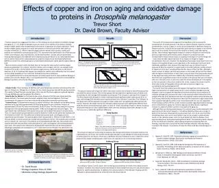
Effects of copper and iron on aging and oxidative damage to proteins in Drosophila melanogaster Trevor Short Dr. David Brown, Faculty Advisor. Introduction. Results. Discussion.

E N D
Effects of copper and iron on aging and oxidative damage to proteins in Drosophila melanogaster Trevor Short Dr. David Brown, Faculty Advisor Introduction Results Discussion Previous research has suggested that there is a link between an accumulation of oxidative damage and aging [1, 2, 3, 4]. Oxidative damage occurs as a result of the inability of the body to detoxify reactive oxygen species that are generated in the body as a byproduct of cellular respiration; These reactive oxygen species may go on to react with proteins in the body and inhibit their ability to function normally [4, 5]. When a reactive oxygen species interacts with a protein, amino acid residues are converted to carbonyl groups [4,5]. This experiment was performed in fruit fliesbecause of their short lifespans, easy maintenance, and the fact that their somatic cells are postmitotic, meaning that damage to the DNA will not be repaired during cell division [1, 2]. Spectrophotometry can be used to measure the carbonyl group by using the compound 2,4-dinitrophenylhydrazine (DNPH) to convert the carbonyl groups to 2,4-dinitrophenylhydrazone, which absorbs light at 380 nm [4,5]. There are factors present within the body that can increase the rate at which reactive oxygen species are generated. For instance, transition metals, such as copper and iron, can catalyze redox reactions that result in the formation of reactive oxygen species, which are known as the Fenton Reactions [3, 4]. It is believed that copper can catalyze this reaction more efficiently than iron based on the relative stabilities of the metal ions involved in the Fenton Reactions. It was hypothesized that an increased exposure to metals would result in more oxidative damage to proteins and a shorter lifespan. Also, it was hypothesized that copper would result in more oxidative damage to proteins and a shorter lifespan. The results of this experiment supported the hypothesis that increasing the concentrations of metals present in the diet can shorten lifespan. However, at lower concentrations, such as 10 ppm, it can not be concluded that a significant impact on lifespan occurred. It should also be noted that acute toxicity to copper or iron did not play a role in the death, as most fruit flies lived for at least 12 days after dosing started. These findings regarding the impact of metals on lifespan are confirmed by previous experiments performed by others. Research has shown that diets contaminated in heavy metals results in a significant decrease in fruit fly lifespan [3]. It was observed that copper had more of an impact on lifespan at higher concentrations than iron. However, the hypothesis that copper would have more of an impact on Drosophila lifespan is not supported, as the results were not found to be statistically significant. Also, because this experiment uses an indirect route of exposure to dose the organism, it would need to be confirmed that a similar amount of copper and iron were actually getting into the tissues of the fruit flies. Although a similar amount of copper and iron were present in the tissues of the fruit flies dosed with the higher concentrations of each metal, only one trial of the body burden aspect of the experiment was performed. Additionally, it should be noted that the reason that there was a significant amount of iron present in the groups that were not dosed with iron is because iron is a necessary metal for normal development in Drosophila, so iron was present in the culture medium [3]. In fact, research has shown that the third instar larvae of Drosophila will actually seek out areas of higher iron concentration during development [3]. The results from the carbonyl assay did support the hypothesis that dosing with higher concentrations of metals would result in more oxidative damage occurring. However, it did not support the hypothesis that there would be an accumulation of oxidative damage that occurred over time, as the carbonyl content measured in the samples after day 20 of dosing was less than the carbonyl content measured after day 10 of dosing. Additionally, the hypothesis that fruit flies dosed with copper would have more oxidative damage to proteins than those dosed with iron was not supported. These findings conflict with the current research available regarding oxidative damage and its relationship to aging. For instance, Agarwal and Sohal found a significant increase in carbonyl content in proteins associated with aging [1]. Although there are discrepancies between the carbonyl assay results observed in this experiment and the current research, it should be noted that there are a couple of factors that may have influenced the results shown here. First of all, more fruit flies should have been used, as 0.3 mg of protein may not be enough to yield accurate results, as the reference paper used 1-4 mg of protein [4]. Also, by 20 days, most experimental groups had experienced significant death. The method by which aliquots of the fruit flies were collected , which was by temporarily paralyzing them via freezing, may have led to some fruit flies that had been dead for a few days being included in the aliquots. The proteins of these fruit flies could have already begun to decay. Lifespan Figure 1: This graph shows the average percent surviving over time for the control group and the experimental groups dosed with copper. Figure 2: This graph shows the average percent surviving over time for the control group and the experimental groups dosed with iron. Methods Lifespan Study- Three replicates of 100 flies each were fed glucose solutions containing either 100 ppm Cu, 10 ppm Cu, 100 ppm Fe, or 10 ppm Fe. The control group was only fed the glucose solution. The number of dead flies was counted every other day and the food supply was replaced regularly. A 2-way ANOVA was used to evaluate the statistics. Body Burden for Metals- For the body burden study, aliquots of 50 flies were dosed using the above methods. The tissues were harvested, weighed, dried, and acid digested. The tissue concentration of copper and iron were determined using atomic absorption spectroscopy. Carbonyl Assay- To determine the amount of protein oxidation, two replicates of the above dosing methods were used with approximately 500 flies being placed in each container. Aliquots of approximately 150 fruit flies were removed after 10 and 20 days. The flies were placed in a dounce homogenizer with pyrophosphate relaxing buffer (PRB) and homogenized and centrifuged. The supernatant was retained and a BCA protein assay was used to determine protein concentrations. Then, 0.3 mg of protein was precipitated using HCl-Acetone (3:100) and the pellet was washed with HCl-Acetone and 10% TCA. The pellet was vortexed during each wash, centrifuged, and the supernatant was discarded. The washed pellets were resuspended in PRB and 10 mM DNPH in 2 M HCl was added. A control sample was treated with just PRB and 2 M HCl. The samples were vortexed every 5 minutes for 0.25 hours. The proteins were precipitated with 30% TCA. The samples were washed with 20% TCA and Ethanol-Ethyl Acetate (1:1), with centrifugation and vortexing between each wash. The samples were dissolved in 6 M guanidine hydrochloride and 20 mM postassium dihydrogen phosphate and incubated in a 37o C water bath and centrifuged. The absorbance was recorded at 380 nm. Figure 3: This graph shows the average percent surviving over time for the control group and the experimental groups dosed with 100 ppm of copper or iron. The groups treated with 10 ppm for either metal had a similar rate of death to that of the group that received the control solution. Flies in these groups did not experience significant rates of death until around day 20, with average 50% mortality occurring day 27 for 10 ppm Cu and day 26 for 10 ppm Fe. The flies treated with 100 ppm for either metal experienced significant death rates around 10 to 12 days, with average 50% mortality occurring around day 13 for 100 ppm Cu and day 17 for 100 ppm Fe. The death rates of flies exposed to 10 ppm for either metal were not significantly different from the control. However, the death rates for the 100 ppm exposure groups were significantly different from both the control and 10 ppm groups (with a p-value of <0.05). According to Figure 3, above, the group dosed with 100 ppm Cu had a slightly higher death rate than the group dosed with 100 ppm Fe, but there was not a statistically significant difference. Body Burden Table 1: This table shows the concentration of metals present in the tissues of the fruit flies after 14 days of dosing for each experimental group. References The amount of exposure to the metal increased as the dosing concentration increased; However, the relationship was not linear. The fruit flies dosed with 100 ppm Cu and 100 ppm Fe had similar amounts of metals present in the tissues. There was a significant amount of exposure to iron in both the control group and the copper-treated groups, which were not exposed to iron in the dosing solutions. • Agarwal S, Sohal RS. 1993. Relationship between aging and susceptibility to • protein oxidative damage. Biochemical and Biophysical Research • Communications .194(3): 1203-1206. • Agarwal S, Sohal RS. 1994. DNA oxidative damage and life expectancy in • houseflies. Proceedings of the National Academy of Sciences of the United • States of America. 91(25): 12332-12335. • 3. Bahadroani S, Hiliker AJ. 2009. Biological and behavioral effects of heavy • metals in Drosophila mealongaster adults and larvae. Journal of • Insect Behavior. 22: 399-411. • 4. Fagan JM, Sleczka BG, Sohar I. 1999. Quantitation of oxidative damage • to tissue proteins. The International Journal of Biochemistry & Cell • Biology. 31(7): 751-757. • 5. Yan LJ, Sohal RS. 1998. Gel electrophoretic quantitation of protein • carbonyl derivativzed with tritiated sodium borohydride. Analytical • Biochemistry. 265(1): 176-182. Carbonyl Assay Death was used as an endpoint. Dead fruit flies were counted every other day. Sample housing container for Drosophila. The 10% glucose solution was placed in the inverted tube, with a piece of cotton being used as a stopper. The food and cotton were replaced regularly. Figure 4: This graph shows the average absorbance measured at 380 nm after 10 days of dosing. Figure 5: This graph shows the average absorbance measured at 380 nm after 20 days of dosing. Acknowledgements According to Figures 5 and 6, above, with increasing concentrations of metals, the carbonyl content increased. However ,the carbonyl content decreased for each sample from day 10 to day 20 (see above figures for specific absorbances). In addition, the carbonyl content of the 100 ppm Fe group and the 100 ppm Cu group appeared to be similar for both day 10 and day 20. • Dr. David Brown • Biology Capstone Class of 2010 • Marietta College Biology Department